
|
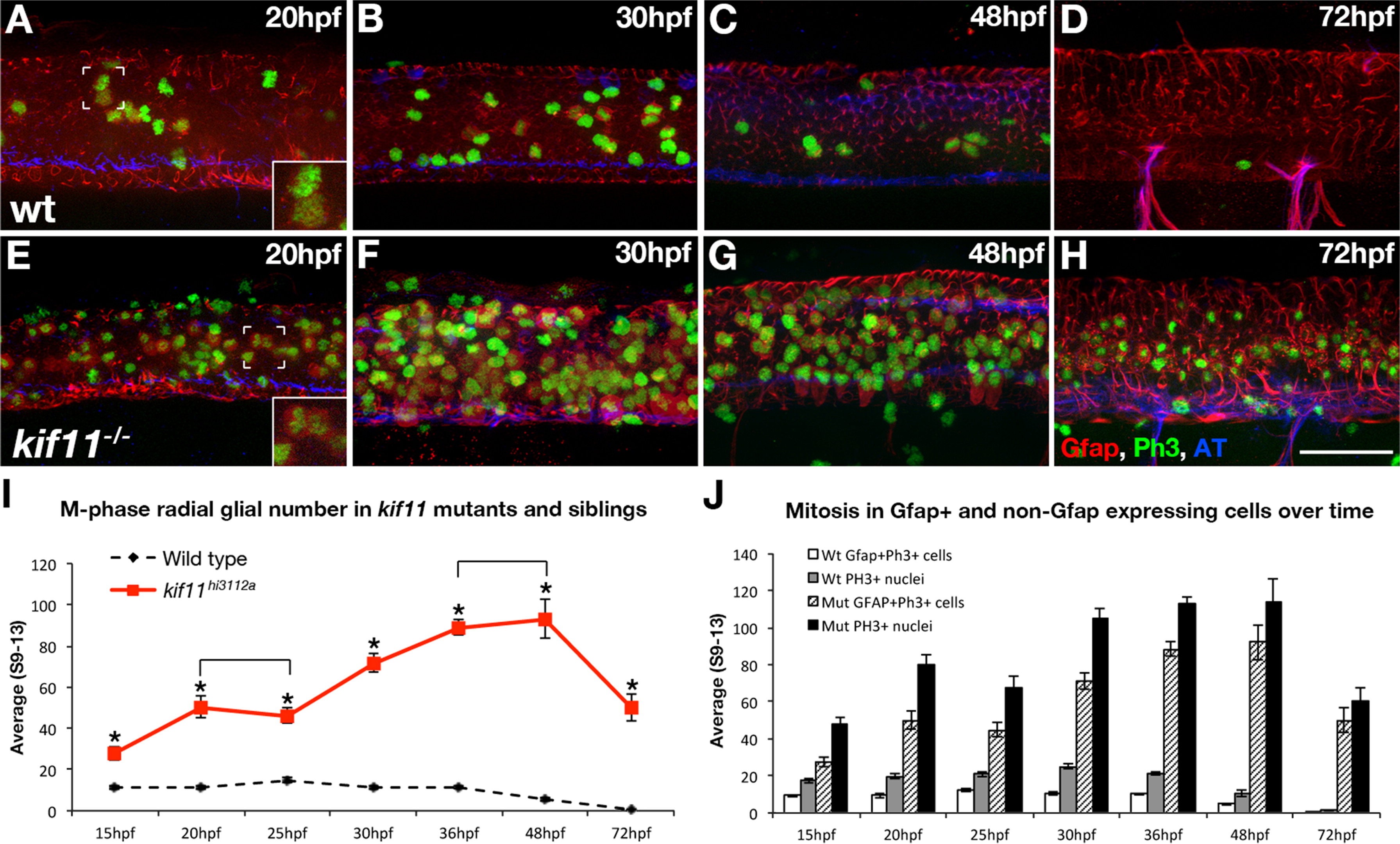
Fig. 2 Loss of kif11 causes an accumulation of radial glia in mitosis. (A–H) Lateral views of kif11-/- sibling neural tubes fixed at timepoints throughout neurogenesis and labeled for radial glia (Gfap), Phosphohistone H3 (PH3) and acetylated Tubulin (AT). Within all siblings, most Gfap+ cells labeled with our antibody were PH3+, indicating these cells were actively proliferating at the time of fixation (A,E insets). At all time points assayed, there were significantly more Gfap+/PH3+ cells in kif11-/- embryos compared with wild type siblings (E–H, quantified in I, asterisks), as well as more PH3+ nuclei (J, shaded and black bars). No change in the number of Gfap+ cells was seen between 20–25 hpf and 36–48 hpf within kif11-/- (I, brackets). Of all proliferating cells (J), it was noted that an unidentified population (PH3+, Gfap) is also kif11 dependent in both wild type and kif11-/- neural tubes. Error bars delineate the standard error of the mean. Asterisks indicate a statistical significance (t-test, two tailed, assuming equal variances, p<0.05).
Reprinted from Developmental Biology, 387(1), Johnson, K., Moriarty, C., Tania, N., Ortman, A., Dipietrantonio, K., Edens, B., Eisenman, J., Ok, D., Krikorian, S., Barragan, J., Golé, C., and Barresi, M.J., Kif11 dependent cell cycle progression in radial glial cells is required for proper neurogenesis in the zebrafish neural tube, 73-92, Copyright (2014) with permission from Elsevier. Full text @ Dev. Biol.