
|
Fig. S4
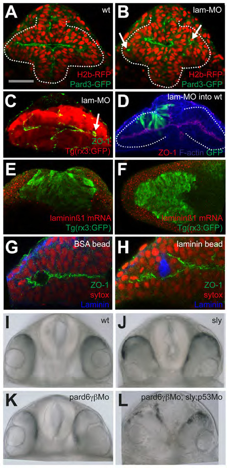
(A-B) wildtype and laminin-γ1 morphants, respectively, expressing H2b-RFP and Pard3- GFP. Arrows in (B) point at mis-positioned pard3-GFP puncta in the eye field of the laminin- γ1 morphant.
(C) laminin-γ1 morphant immunostained as detailed in the panel. Arrows point at mispositioned ZO-1 puncta.
(D) laminin-γ1 morphant cells (green) transplanted into a wild type eye field show no overt phenotype. Embryo immunostained as detailed in the panel.
(E-F) frontal and dorsal views, respectively, of 3ss Tg(rx3:GFP) embryos showing laminin-β1 mRNA accumulation (red) and GFP protein (green). laminin-α1 and laminin-γ1 show similar expression patterns (not shown).
(G-H) BSA-coated (G) and Laminin1-coated (H) beads implanted in the eye field. Embryos were left to develop until 10ss and immunostained as detailed in the panels.
(I-L) Frontal views of 24hpf embryos of the genotypes detailed in the panels.
Reprinted from Developmental Cell, 27(3), Ivanovitch, K., Cavodeassi, F., and Wilson, S.W., Precocious acquisition of neuroepithelial character in the eye field underlies the onset of eye morphogenesis, 293-305, Copyright (2013) with permission from Elsevier. Full text @ Dev. Cell