
|
Fig. 4
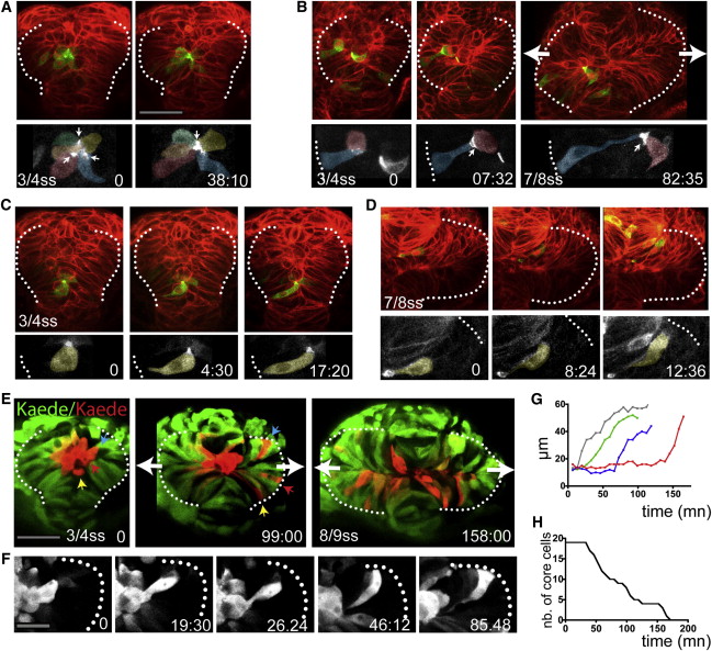
Core Eye Field Cells Polarize and Intercalate in the Marginal Layer during Eye Morphogenesis
(A–D) Time-lapse movies of eye field core cells expressing the fusion protein Pard3-GFP (green). Embryos are counterlabeled with a membrane-RFP (red). Insets below show the green channel only (Pard3-GFP).
(E) Time-lapse movie of an embryo expressing Kaede. Core cells were labeled in red by photoconversion of Kaede from green to red. Colored arrows point at individual core cells as they intercalate within the optic vesicles.
(F) Close-up of a single core cell elongating and acquiring a wedge shape as it intercalates.
(G) Quantification of cell length over time in individual core cells with data extracted from the movie sequence in (E).
(H) Graph showing the reduction in the number of core cells as morphogenesis proceeds (18 core cells were tracked from t = 0 onward).
White dotted lines in (A)–(F) outline the eye field. Lateral arrows in (B) and (E) indicate the direction of evagination of the tissue. Scale bars, 64 μm (A and E) and 27 μm (F). See also Figure S3 and Movies S4, S5, S6, S7, and S8.
Reprinted from Developmental Cell, 27(3), Ivanovitch, K., Cavodeassi, F., and Wilson, S.W., Precocious acquisition of neuroepithelial character in the eye field underlies the onset of eye morphogenesis, 293-305, Copyright (2013) with permission from Elsevier. Full text @ Dev. Cell