
|
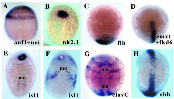
Fig. 5 Noggin-injected embryos are more severely dorsalised than swr- embryos. Lateral (A), animal pole (B-D) and dorsal (E-H) views of of 1-2 somite stage (except A, 90% epiboly) embryos injected with noggin RNA. (A) anf and noi expression is radialised in noggin-injected embryos (similar to swr- embryos; compare with Figs 1D and 2H). (B) nk2.1 expression is retained in the prospective hypothalamus of the injected embryo. (C,D) Marginal neural plate expression of flh, emx1 and fkd6 is lost in noggin-injected embryos (compare with Fig. 2D,F). (E) Mild and (F) massive expansion of isl1 expressing motor neurons in noggin-injected embryos. The asteriks indicates the pillow. (G) Radial expansion of elavC expressing medial neurons in a severely dorsalised noggin-injected embryo. (H) shh expression in axial tissue in a noggin-injected embryo. me, medial neurons; mn, motor neurons.