
|
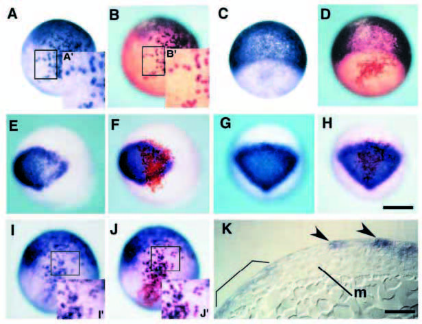
Fig. 7 Constitutively active BMPR-IA can simulate BMP signaling in a direct and cell-autonomous manner in ectoderm patterning. (A-J) Chimeras containing cells derived from injected embryos with biotinylated dextran as a lineage tracer and mRNA encoding either constitutively active receptor (A,B, E,F, I,J) or b-galactosidase (C,D, G,H). Embryos after operation are processed for whole-mount in situ hybridization at 80% epiboly with gata-3, epidermal marker gene (A,C), otx-2, neural marker gene (E,G) and zbmp-2 (I). The same embryos are stained with avidin-biotin HRP complex for the purpose of detection of donor cells (B,D), (F,H), (J), respectively). The insets (A′,B′, I′,J′) show a magnification of the boxed area in the same figures. Note that BMP signaling can activate the expression of gata-3 and zbmp-2 only in cells derived from injected embryo (compare Fig. 7A′ with 7B′ and 7I′ with 7J′, respectively). By contrast, otx-2 is downregulated only in donor cells expressing CABRIA (E,F). In chimeric embryos transplanted cells containing b-galactosidase (C,D, G,H), neither ectopic induction nor repression of marker genes were detected. (K) Sagittal section of chimeric embryos containing CA-BRIA in ectoderm. Ectopic gata-3 signals are observed in ectoderm (arrowheads), suggesting that ectoderm ventralization is independent of mesoderm ventralization. Endogenous gata-3 expression is marked by bracket. Abbreviation: m, axial mesoderm. Scale bar, 200 μm (H), 50 μm (K). A-J are the same magnification.