
|
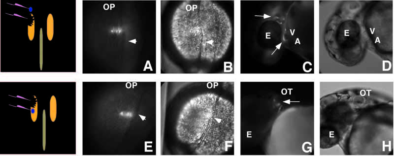
Fig. 5 Cells that replace cardiac progenitors arise anterior to the position of normal cardiac progenitors. Fluorescent (A,C,E,D) and phase (B,D,F,G) images of cell labeling (blue dot) after ablation in 10-somite-stage embryos. The schematic images indicate the location of the ablation and cell labeling in relation to the notochord and Nkx2.5 expression. (A,B) Labeled fluorescent cells anterior to the ablation of the heart precursors give rise to cells in both the cranial mesenchyme and the heart (arrows; C,D). The purse-string-like contraction of the tissue surrounding the ablation have caused the labeled cells to elongate. (E,F) In contrast, labeled cells posterior to the ablation contribute to the mesenchyme around the otic vesicle but not the heart (arrow; G,H). For orientation, the optic cup (OP), eye (E), otic vesicle (OT), ventricle (V) and atrium (A) are labeled. Images are labeled as described in Fig. 1.