
|
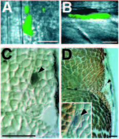
Fig. 8 Adaxial cells become slow muscle fibers. (A) Dorsal view of the segmental plate of an approximately 12 h embryo (6 somites), after injection of adaxial cells with lysinated fluorescein dextran. Two or three adaxial cells were injected in this embryo, acellular fluorescent debris is present lateral to the injected adaxial cells. (B) Side view of the same embryo at 8 days. Only one of the injected adaxial cells survived. It developed into a muscle cell located in somite 11. The hexagonal background pattern in this image is due to the video camera. (C) Transverse section of the same embryo at 20 d. The section has been labeled with an antibody to fluorescein, thus the injected cell is black (arrowhead). (D) The same section after labeling with the 12/101 antibody. The adaxial cell developed into a small diameter muscle fiber located in the slow muscle region, not labeled by 12/101 (arrowhead). Inset: higher magnification (as in C) view of the injected cell (arrowhead). In a series of similar experiments, 4 out of 4 injected adaxial cells became slow muscle fibers, as determined by antibody staining, position and morphology. In dorsal views, anterior is up; in side views, anterior is to the left; in transverse sections, dorsal is up. Scale bars, 50 μm.