
|
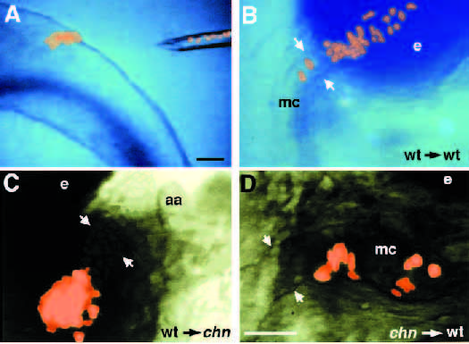
Fig. 5 Mutant neural crest cells form cartilage in the presence of wild-type neighbors. (A) 12 hour, lateral view. Neural crest cells from a donor embryo previously marked with lineage tracer dye are shown after transplantation into the premigratory neural crest of an unlabeled host. A number of labelled cells remain in the suction micropipette. (B) 72 hour, ventral view. In a control, wild-type neural-crest-derived cartilage is identifiable by its morphology in Meckel’s cartilage and the palatoquadrate in a wild-type host. (C) 72 hour, lateral view. A mosaic embryo made by transplanting wild-type neural crest into a chn host. Labelled cartilage cells from the wild-type donor are visible as well as unlabelled cartilage derived from the mutant host (between arrows). (D) 72 hour, ventral view. A mosaic consisting of mutant neural crest transplanted into a wild-type host also shows labelled and unlabelled cartilage. Abbreviations: aa, aortic arch; e, eye; mc, Meckel’s cartilage. Scale bars, 100 μm.