
|
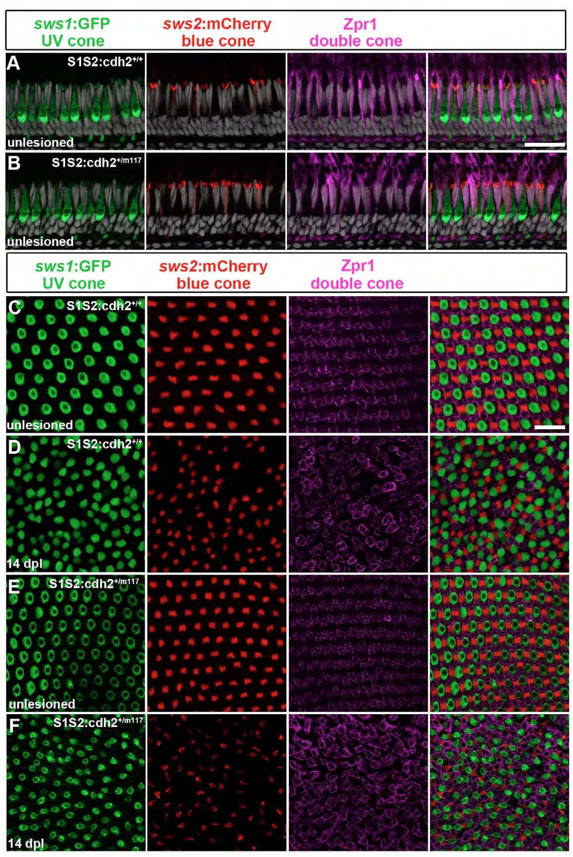
Fig. S5 Regeneration of cone photoreceptors in Tg(sws1:GFP;sws2:mCherry); cdh2+/m117 heterozygote retinas. (A, B) Immunocytochemistry for red/green double cone marker, zpr1 (magenta); UV cones (green), blue cones (red). In unlesioned retinas, all cone subtypes are present and appear morphologically normal in the cdh2+/m117 het retinas (B) similar to the wild-type sibs (A). (C-F) Immunocytochemistry for zpr1 (magenta) in unlesioned, flat-mounted retinas illustrates the organized cone mosaic pattern in wild-type sib (C) and cdh2+/m117 het retinas (E). At 14 dpl, all cone subtypes regenerate, but the mosaic pattern is not restored in sib (D) or cdh2+/m117 (F). Scales: 20 μm, A-F.