
|
Fig. S3
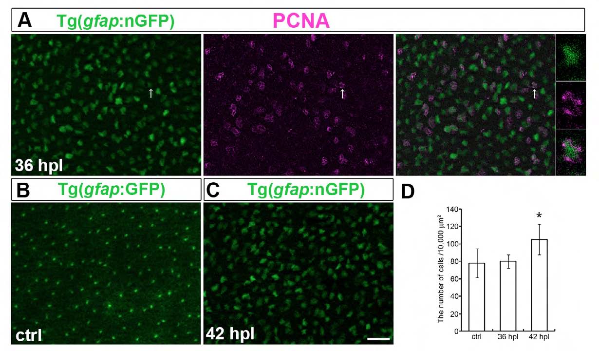
Injury-induced Muller glia reenter the cell cycle and complete cell division between 36 and 42 hpl.
(A) PCNA (magenta) immunocytochemistry on flat-mounted retina from a light lesioned mi2004 fish with inducible nGPF (green) at 36 hpl; some nGFP+ Muller glia express PCNA (arrow). (B) Flat-mounted, unlesioned retina from mi2002 fish with gfap:GFP reporter, focused at the level of the basal processes of Müller glia (green) in the inner plexiform layer. (C) Müller glia in a lightlesioned mi2004 fish with inducible nGFP (green) in at 42 hpl. (D) Planimetric density of GFP+ Müller glial cells. Scales: 20 μm A, B, C. * p < 0.005.