
|
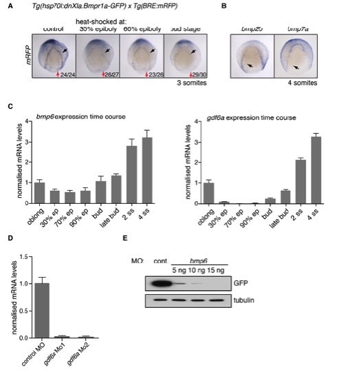
Fig. S1 Identification of the BMP/GDF ligands involved in BMP signalling at the NPB. (A) ISH for mRFP on Tg(hsp70l:dnXla.Bmpr1a-GFP) x Tg(BRE:mRFP) embryos, heat-shocked at the indicated stages, demonstrates inhibition of BMP signalling. Black arrows indicate outer domains and red arrows indicate inner domains of BMP activity. (B) ISH for bmp2b and bmp7a on wild-type embryos. Black arrow indicates the position of neural crest at this developmental stage. (C) Temporal expression analysis of bambi-b, and gdf6a using qRT-PCR. The levels were normalised to those of ef1a. Developmental stages are indicated. (D) qRT-PCR indicates that the splice-blocking gdf6a MO1 and MO2 inhibit splicing of gdf6a exon1 to exon2. The levels were normalised to those of ef1a. (E) The bmp6 MO is effective as a translation blocker. The bmp6 MO binding site was cloned upstream and in frame with GFP in pCS2+. The plasmid was transcribed and translated in rabbit reticulocyte lysate in the presence of increasing amounts of bmp6 MO, the concentrations of which corresponded to in vivo doses of between 5 and 15 ng per embryo. GFP production was assayed by Western blot and tubulin acts as a loading control. In A the numbers indicate embryos with the observed phenotype for a representative experiment.