
Fig. S1
- ID
- ZDB-IMAGE-140107-36
- Publication
- Fernandes et al., 2013 - Orthopedia Transcription Factor otpa and otpb Paralogous Genes Function during Dopaminergic and Neuroendocrine Cell Specification in Larval Zebrafish
- All Figures
- Figures for Fernandes et al., 2013
|
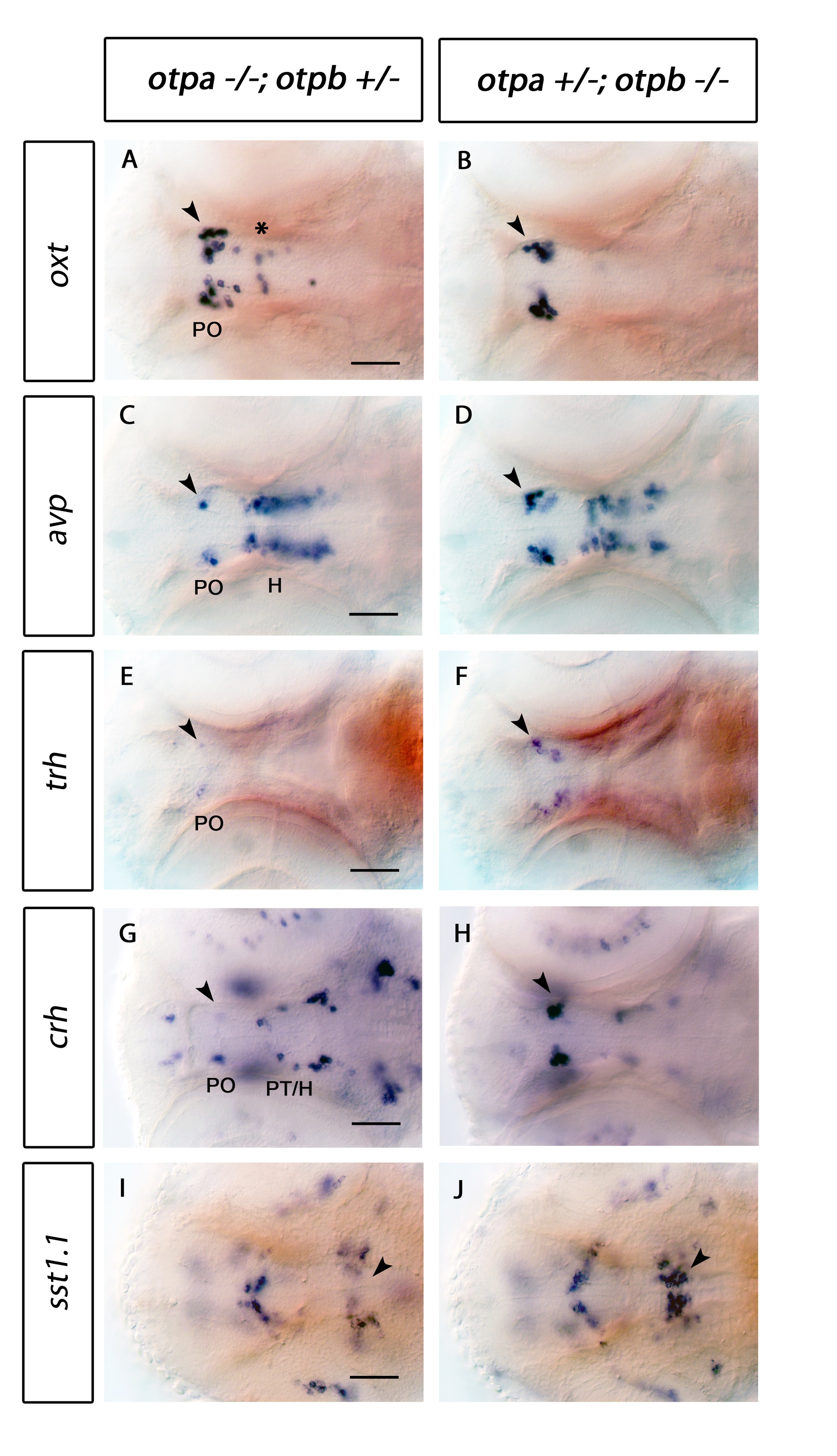
Fig. S1 Expression of oxt, avp, trh, crh and sst1.1 in otpa and otpb mutant larvae. Whole-mount in situ hybridization of 3 dpf larvae reveals changes of oxt, avp, trh and crh expression in the preoptic region (arrowhead in A, C, E, G) and reduction of sst1.1 expression (arrowhead in I) in the hindbrain of otpa-/- mutant, otpb+/- heterozygous larvae. In contrast, no obvious change is detected in the preoptic region (arrowheads in B, D, F, H) and hindbrain (J) of otpb-/- mutant, otpa+/- heterozygous larvae. Dorsal view, anterior at left. Scale bar is 50 μm. H, hypothalamus; PO, preoptic region; PT, posterior tuberculum.