
Image
|
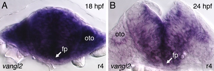
Figure Caption
Fig. S1
Vangl2 is expressed broadly in the hindbrain and surrounding tissues during the period of FBM neuron migration.
(A,B) Cross sections (30 microns thickness) in rhombomere 4 of 18 and 24 hpf zebrafish hindbrains processed for vangl2 wholemount in situs (purple) reveals expression in the neural tube, and adjacent mesendoderm. fp, floor plate; oto, otocyst.
Acknowledgments
This image is the copyrighted work of the attributed author or publisher, and
ZFIN has permission only to display this image to its users.
Additional permissions should be obtained from the applicable author or publisher of the image.
Reprinted from Developmental Biology, 382(2), Sittaramane, V., Pan, X., Glasco, D.M., Huang, P., Gurung, S., Bock, A., Li, S., Wang, H., Kawakami, K., Matise, M.P., and Chandrasekhar, A., The PCP protein Vangl2 regulates migration of hindbrain motor neurons by acting in floor plate cells, and independently of cilia function, 400-412, Copyright (2013) with permission from Elsevier. Full text @ Dev. Biol.