
|
Fig. 1
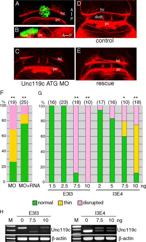
Unc119c is involved in HC formation in zebrafish embryos. A, B: Visualizing the HC. Transgenic zebrafish, Tg(AANAT2:eGFP), which express eGFP in the pineal gland, were used. The embryos were fixed at 54 hpf and stained with anti-GFP (green, pineal gland) and anti-acetylated tubulin (red, axonal tracts). The HC is located directly beneath the pineal gland. Dorsal view (A), lateral view (B). C–G: Injection of unc119c MO disrupts HC formation. Wild type zebrafish embryos were injected with 1.5 ng unc119c ATG MO plus 100 pg GFP RNA (C, F), control MO (D), or unc119c ATG MO together with 100 pg unc119c RNA (E, F). Two different unc119c splice MOs, E3I3 and I3E4, also affected HC formation in a dose-dependent manner (G). Statistical significance is indicated as *P < 0.05, and **P < 0.01. Experiments are compared to HC in control embryos (all of which were normal), except for the rescue data (F, second bar, MO+RNA), which are compared to MO-injected embryos (F, first bar). Total number of embryos examined is shown in parentheses above the bars. H: The splice MOs greatly reduced formation of mature mRNA. dvdt, dorsoventral diencephalic tract; hc, habenular commissure; p, pineal gland; pc, posterior commissure.