
Image
|
Figure Caption
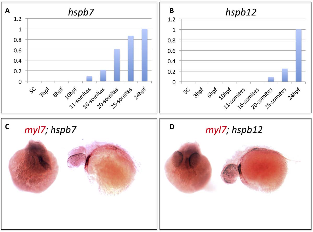
Fig. S3
Expression patterns of hspb7 and hspb12 transcripts in the embryonic heart. A, B) qRT-PCR results measuring relative expression levels for hspb7 and hspb12 transcripts during early zebrafish embryogenesis. C, D) In situ hybridization analysis showing representative 48 hpf embryos stained with probes for the cardiac marker myl7 (red) and hspb7 or hspb12 (black). The stains overlap in the embryonic heart, shown in either head-on (left) or anterior-left (right) views.
Acknowledgments
This image is the copyrighted work of the attributed author or publisher, and
ZFIN has permission only to display this image to its users.
Additional permissions should be obtained from the applicable author or publisher of the image.
Reprinted from Developmental Biology, 381(2), Rosenfeld, G.E., Mercer, E.J., Mason, C.E., and Evans, T., Small heat shock proteins Hspb7 and Hspb12 regulate early steps of cardiac morphogenesis, 389-400, Copyright (2013) with permission from Elsevier. Full text @ Dev. Biol.