
|
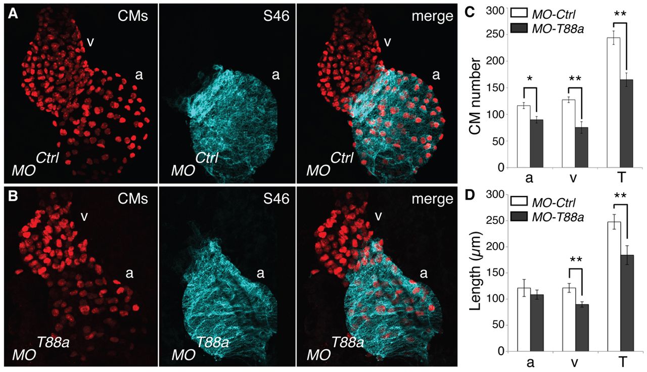
Fig. 5
Tmem88a-deficient hearts are small and hypocellular. (A,B) Representative confocal images of control and tmem88a morphant hearts dissected from 51 hpf zebrafish embryos. Morphants were generated in the tg(myl7:DsRed2-nuc) background. Left panels show immunohistochemistry for DsRed2 labeling of cardiomyocyte nuclei (CMs). Middle panels show S46 antibody labeling of atrial cells. Right panels show merge. Atrium (a) and ventricle (v) are indicated. (C) Atrial (a), ventricular (v) and total (T) cardiomyocyte numbers. (D) Atrial, ventricular and total cardiac chamber length. n=6 hearts quantified per condition. *P<0.05, **P<0.01; error bars indicate s.d.