
Image
|
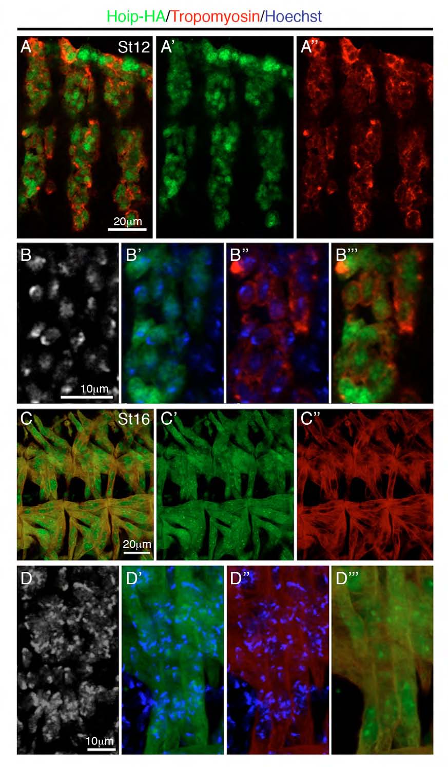
Figure Caption
Fig. S5
Hoip localizes to both the nucleus and the cytoplasm. (A-D) Mef2>Hoip-HA embryos co-labeled for HA (green) and Tropomyosin (red). (A,B) Hoip is largely localized to the nucleus in St12 embryos, although some cytoplasmic staining is present. (C,D) Hoip is localized throughout the myofibers of St16 embryos. Enhanced localization is apparent in a subnuclear domain.
Acknowledgments
This image is the copyrighted work of the attributed author or publisher, and
ZFIN has permission only to display this image to its users.
Additional permissions should be obtained from the applicable author or publisher of the image.
Full text @ Development