
|
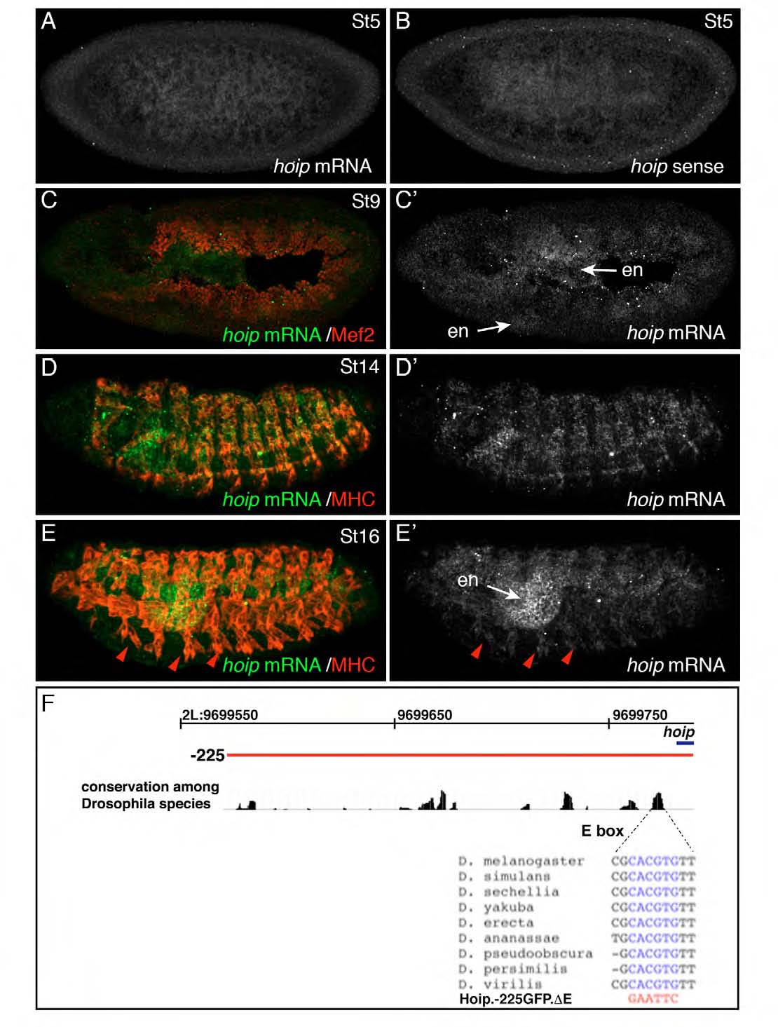
Fig. S4
hoip expression during embryonic development. (A-E) Wild-type embryos hybridized with RNA probes antisense (A,C-E) or sense (B) to the hoip mRNA. (A,B) Post-cellularization blastoderm embryos. Fluorescent intensity is comparable between the antisense and sense probes. (C) St9 embryo co-labeled for hoip (green) and Mef2 (red). Weak hoip expression has initiated in the endoderm and mesoderm. (D,E) St14 (D) and St16 (E) embryos co-labeled for hoip (green) and MHC (red). hoip is expressed at high levels in the endoderm (en) and at lower levels in the somatic musculature (red arrowheads). (F) Genomic conservation 225 bp 52 to the hoip transcriptional start site. The highly conserved E-box sequence and mutations in Hoip.-225.DEGFP are shown. Genomic coordinates refer to base pair positions along chromosome 2L.