
Image
|
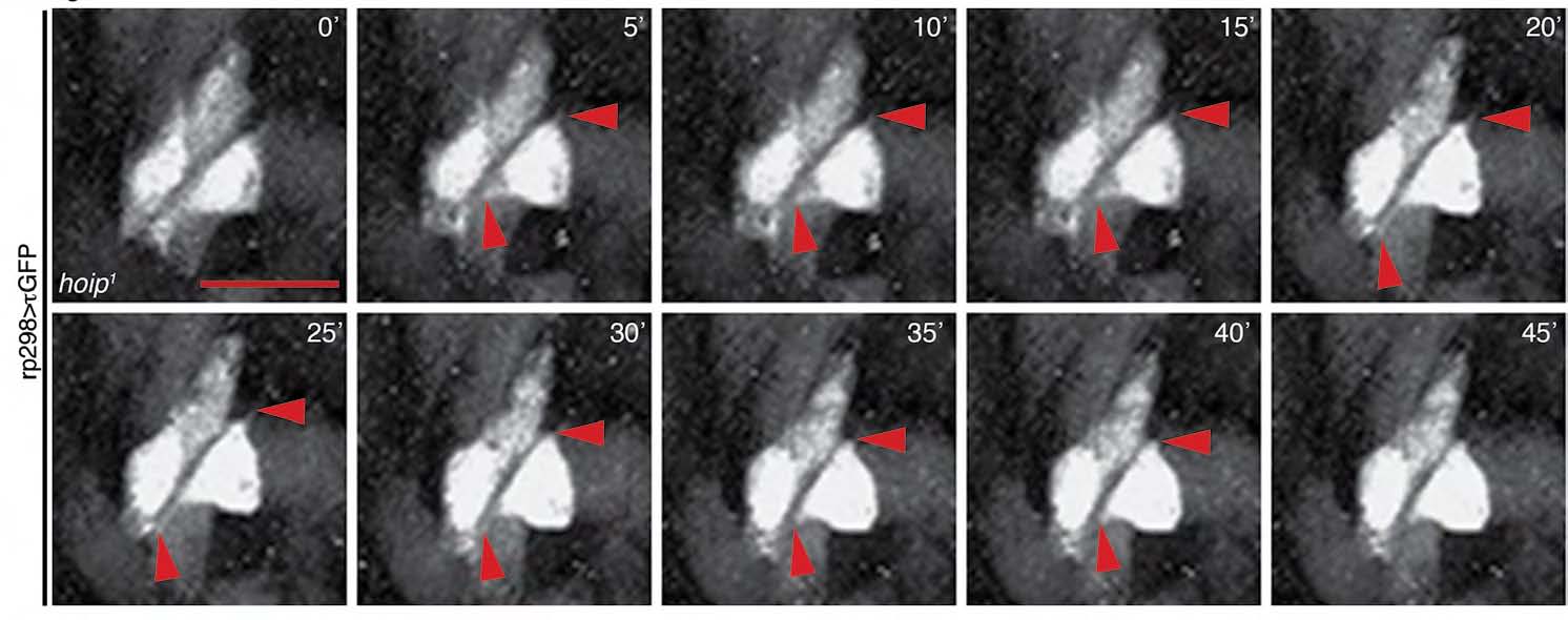
Figure Caption
Fig. S2
Somatic muscles in hoip embryos extend filapodia in the direction of polarization. Time-lapse images of rp298. gal4>τGFP hoip1 somatic muscles beginning at late St12. Filapodia extend in the axis of myotube polarization (red arrowheads) and then retract. Scale bar: 10 μm.
Acknowledgments
This image is the copyrighted work of the attributed author or publisher, and
ZFIN has permission only to display this image to its users.
Additional permissions should be obtained from the applicable author or publisher of the image.
Full text @ Development