
|
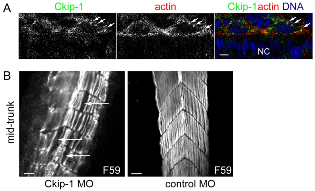
Fig. 3 Ckip-1 knock down does not affect slow muscle cell migration through the fast myotome. (A) Confocal image of adaxial cells at the depth of the notochord in 8- to10-somite embryos (14 hpf) labelled with Ckip-1 (green), phalloidin (red) and DAPI (blue). Dorsal view. Arrows indicate Ckip-1 colocalisation with actin. NC: notochord. Scale bar: 5µm. (B) Single frames of a 3D reconstitution encompassing a five-somite width lateral view of 48hpf Ckip-1-MO- and control-MO-injected embryos labelled with F59 antibody (grey) that stains slow fibres. Anterior to the top. Arrows indicate gaps in the lateral palisade of slow muscle fibres. Scale bars: 25µm.