
|
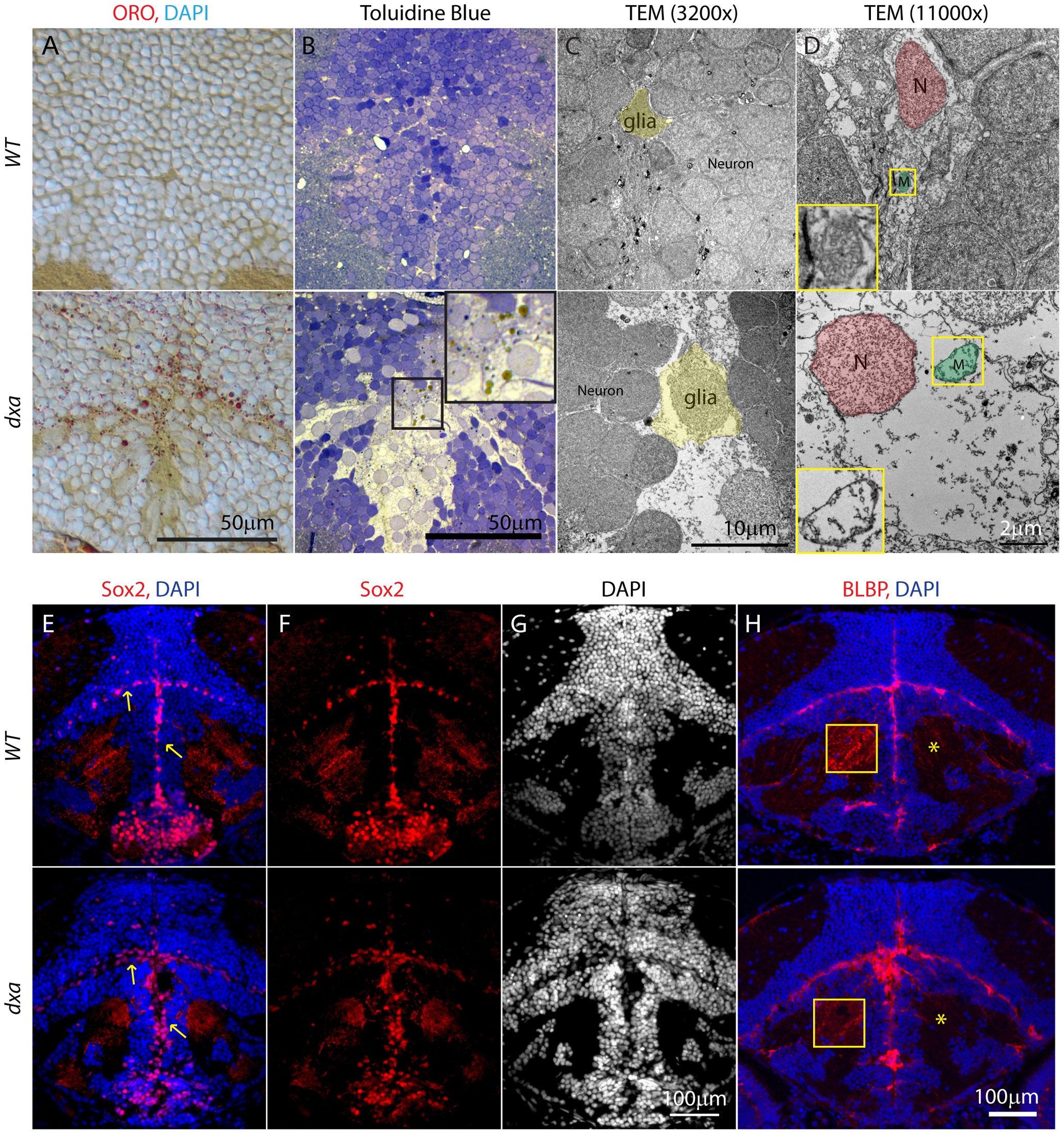
Fig. 4
Lipid accumulation and necrotic features in the dxavu463 mutants with increased numbers of neural progenitor cells and dysmorphic brain.
Top panels show control wild type zebrafish with bottom panels showing images from type II dxa mutant at 8 dpf. (A) ORO (red) showing increased lipids in the mutant brain, DAPI (bright blue) staining nuclei. (B) Toluidine blue staining in wild type and type II dxa with severe brain defects. Magnified view of midline region in the rectangle is shown on the upper right corner with greatly enlarged neural progenitor cells and neurons. Brown colored vesicles again suggest lipid drops containing cerebroside sulfate. (C) TEM image of VZ in WT (top) and type II dxa (bottom). Yellow pseudocolor indicates a single glia cell in WT and dxa mutants, marked increase in cell size is present. (D) Higher magnification image of neural progenitor cells. Green pseudocolor region indicates individual mitochondria. Pseudocolor with red indicates nuclei. Magnified views of normal and mutant mitochondria are shown on the left lower bottom. (E) Anti-Sox2 (red) and DAPI (blue) staining in control (top) and dxa zebrafish (bottom). Yellow arrows indicate Sox2 positive cells in the VZ. (F) Red channel image of (E). (G) DAPI channel of (E) showing disrupted gray matter of brain. (H) Anti-BLBP staining in wild type (top) and dxa (bottom). Asterisks indicate white matter region normally containing glia fibers. Region within the yellow box is further magnified to allow fine details of glial fibers to be seen. Contrast levels of control (top) and dxa zebrafish (bottom) were adjusted together to compare glial fibers. Scale bar = 50 μm (A, B), 10 μm (C), 2 μm (D) and 100 μm (E–H).