
|
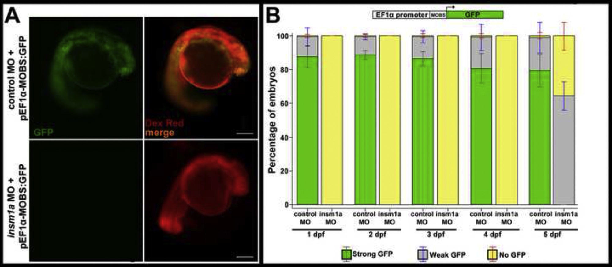
Fig. S1
The insm1a MO is highly effective through 4 dpf. Embryos were co-injected with either standard control or insm1a MO and an eF1α-GFP plasmid containing the insm1a MO binding site (pEF1α-MOBS:GFP; n>40 embryos for each group). (A) At 1 dpf, control MO injected embryos showed robust, ubiquitous expression of GFP (top panels), whereas insm1a MO injected embryos showed no expression of GFP (bottom panels). Tetramethylrhodamine dextran (Dex Red) was used as an internal injection control. (B) Quantitation of the numbers of injected embryos showing GFP expression from 1 dpf through 5 dpf. GFP expression similar to that shown in (A) (top panel) was categorized as “strong GFP”. GFP expression that was limited to just a few cells in the embryo was categorized as “weak GFP” expression. “No GFP” required the complete absence of any identifiable GFP signal. From 1 dpf through 4 dpf, no insm1a morphants exhibited any GFP expression. In contrast, nearly all embryos co-injected with the standard control MO were GFP-positive, with over 90% strongly expressing GFP. By 5 dpf, nearly half of the insm1a morphants showed low levels of GFP expression, indicating a decline in the effectiveness of the insm1a MO. Scale bar=200 μm; MOBS, insm1a morpholino binding site; MO, morpholino; dpf, days post fertilization.
Reprinted from Developmental Biology, 380(2), Forbes-Osborne, M.A., Wilson, S.G., and Morris, A.C., Insulinoma-associated 1a (Insm1a) is required for photoreceptor differentiation in the zebrafish retina, 157-171, Copyright (2013) with permission from Elsevier. Full text @ Dev. Biol.