
|
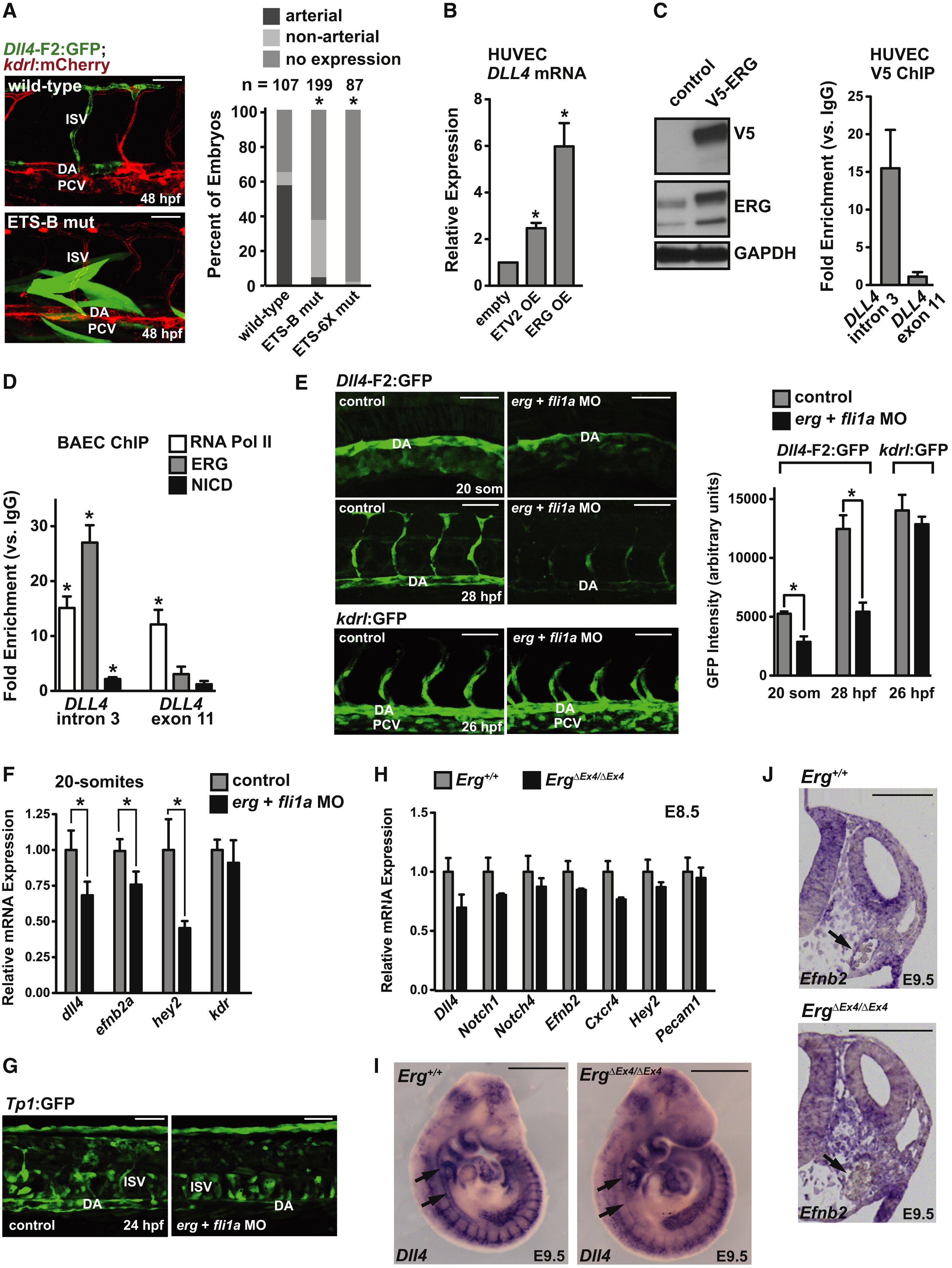
Fig. 4
Dll4 Is Regulated by ETS Factors (A) Representative images (left) and quantification (right) of wild-type, ETS-B, and ETS-6x mutant F2:GFP embryos at 48 hpf. ISV, intersomitic vessel; DA, dorsal aorta; PCV, posterior cardinal vein. Asterisk indicates a significant difference in arterial expression compared to wild-type (χ2 test). (B) Expression of endogenous DLL4 in HUVEC electroporated with ETV2 or ERG expression constructs (n = 3, ETV2; n = 5, ERG). (C) V5-ERG expression in electroporated HUVEC (left). ChIP in V5-ERG-electroporated HUVEC. Fold enrichment (V5 versus IgG) was measured at the Dll4 enhancer (intron 3) and Dll4 exon 11 (n = 2). (D) ChIP in BAEC for RNA Polymerase II (RNA Pol II), ERG or the Notch intracellular domain (NICD) (n = 3). Asterisk indicates significant enrichment over IgG control. (E) Expression of F2:GFP (arterial) and kdrl:GFP (pan-endothelial) in erg;fli1a morphants. Representative images (left) and quantification (n = 5–10) (right). (F) Endogenous dll4, ephrinb2a, hey2, and kdr in control and erg;fli1a morphants assessed by qPCR (n = 5–10 individual embryos). (G) Notch activity (Tp1:GFP) was diminished in the dorsal aorta of <30% of erg/fli1a morphants. (H) Levels of arterial markers and a pan-endothelial marker (Pecam1) were quantified by qRT-PCR in E8.5 embryos (n = 3 for wild-type, n = 2 for ErgΔEx4/ ΔEx4 embryos). (I) Endogenous Dll4 mRNA is downregulated in the DA of ErgΔEx4/ ΔEx4 embryos (arrows; n = 3 embryos per genotype) at E9.5. (J) Efnb2 section in situ hybridization at 9.5 shows downregulation of Efnb2 in the DA (arrows) of ErgΔEx4/ ΔEx4 embryos (Erg+/+, n = 3; ErgΔEx4/ ΔEx4 n = 2). Scale bars represent 50 μm (A, E, and G), 500 μm (I), 100 μm (J). All graphical data are mean ± SEM. See also Figures S3 and S4.
Reprinted from Developmental Cell, 26(1), Wythe, J.D., Dang, L.T., Devine, W.P., Boudreau, E., Artap, S.T., He, D., Schachterle, W., Stainier, D.Y., Oettgen, P., Black, B.L., Bruneau, B.G., and Fish, J.E., ETS Factors Regulate Vegf-Dependent Arterial Specification, 45-58, Copyright (2013) with permission from Elsevier. Full text @ Dev. Cell