
|
Fig. 6
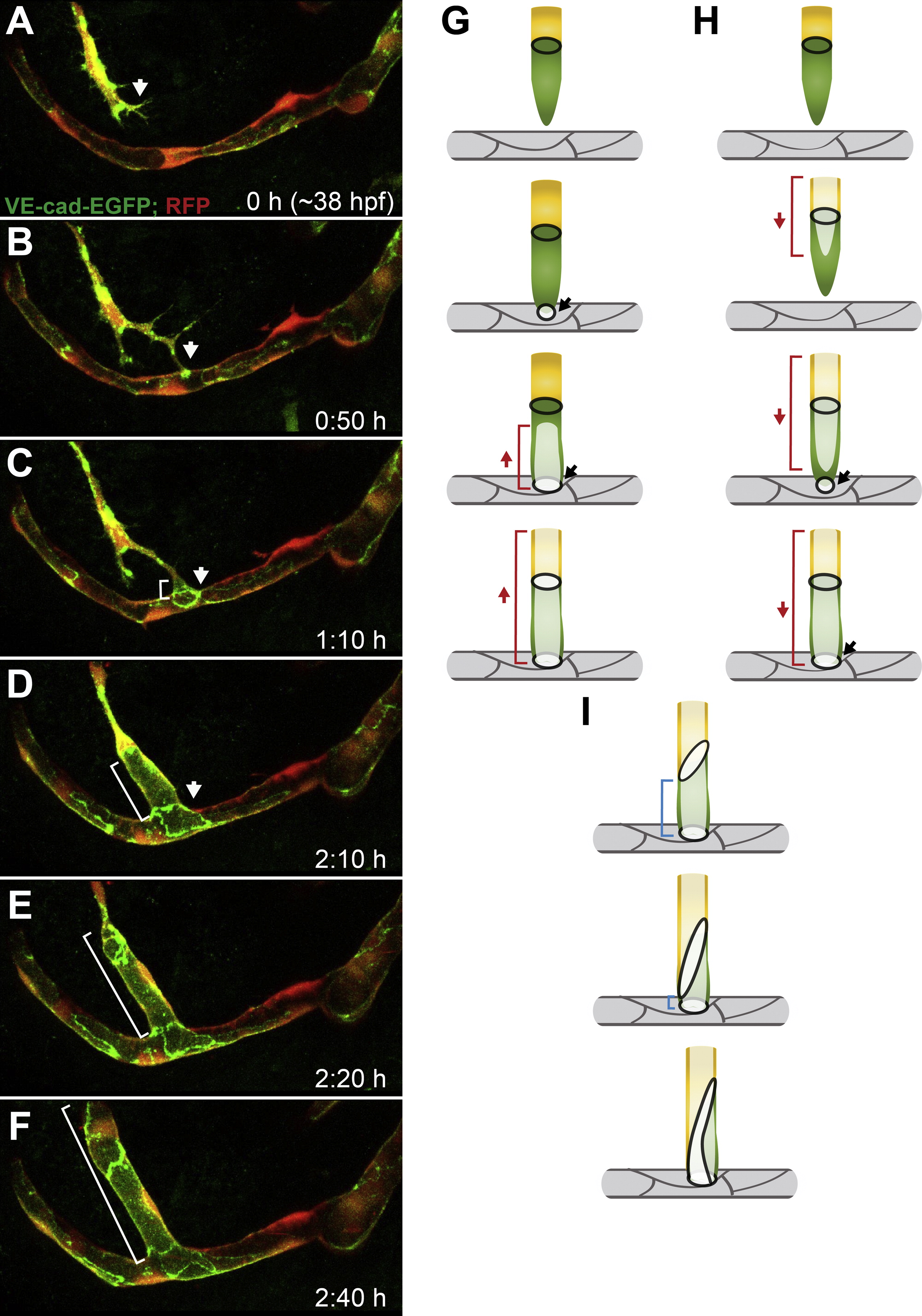
Fusion of a CMV Sprout to an Existing PLA Vessel (A–F) Still pictures of a time-lapse movie showing fusion of CMV to PLA in a transgenic embryo Tg(fliep:GFF)ubs3,(UAS:mRFP),(UAS:VE-cadherinΔC-EGFP)ubs12. CMV (communicating vessel) sprouts ventrally toward the PLA (A, arrow). The leading tip cell connects to the PLA on the cell body of one of the ECs making a spot of junctions (B, arrow). The newly formed ring connects to an existing junctional line on the PLA (C, arrow) and transcellular lumen forms in the CMV tip cell from the PLA upward (C, white bar). The lumen extends through the whole sprout (E and F, white bar). (G) A cellular model of the CMV-PLA fusion. The CMV tip cell is green, followed by a yellow stalk. The PLA is bright gray with dark gray junctions. CMV makes a new junctional connection to the PLA (black ring, arrow). Lumen (bright green, red bar) is pushed through the tip cell and inflates up the sprout. (H) A model of an alternative lumen formation process, where the CMV is lumenized from the CMV sprout toward the PLA (follow the red bars). (I) Blue bars mark the cellular/junctional rearrangements leading to transformation of the CMV into a multicellular tube. See also Movie S6. Scale bars, 20 μm.
Reprinted from Developmental Cell, 25(5), Lenard, A., Ellertsdottir, E., Herwig, L., Krudewig, A., Sauteur, L., Belting, H.G., and Affolter, M., In Vivo analysis reveals a highly stereotypic morphogenetic pathway of vascular anastomosis, 492-506, Copyright (2013) with permission from Elsevier. Full text @ Dev. Cell