
|
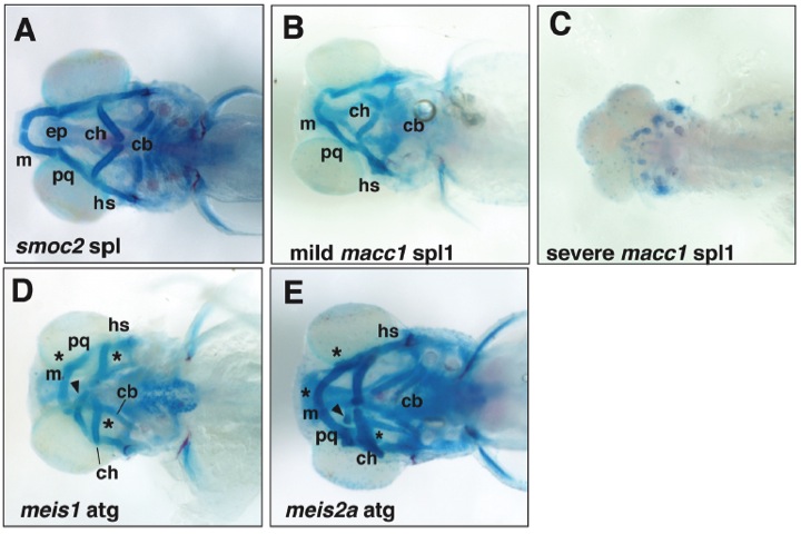
Fig. S6 Examples of morphant phenotypes maintained in the absence of p53. All images are of the morphant phenotypes seen in p53 null larvae. A, Ventral view of viscerocranial cartilages of a 5dpf smoc2 SPL morphant showing inversion of the ceratohyal, and a reduction in ceratobranchial size and number. Figure 2 shows similar morphant phenotype obtained in wild-type larva. B, C, Compare with macc1 morphant phenotypes obtained in wild-type larva shown in Figure 5. B, Ventral view of viscerocranial cartilages of a 5dpf macc1 spl1 morphant larva presenting with a mild phenotype - loss of ceratobranchials, inversion of the angle between the paired ceratohyal and reduction of Meckel’s cartilage. C, Ventral view of viscerocranial cartilages of a 5dpf macc1 SPL1 morphant presenting with a severe phenotype – almost complete aplasia of all head cartilages. D, E, Compare with meis1 and meis2a morphant phenotypes obtained in wild-type larva shown in Figure 3. D, Ventral view of viscerocranial cartilages of a 5dpf meis1 ATG morphant larva showing cartilage fusions between Meckel’s and palatoquadrate cartilages and ceratohyal and ceratobranchial cartilages. E, Ventral view of viscerocranial cartilages of 5dpf meis2a ATG morphant larva showing cartilage fusions between paired Meckel’s cartilages, Meckel’s and palatoquadrate, and ceratohyal and ceratobranchial cartilages. Both meis morphants also show ectopic cartilages (arrowhead) and cartilage fusions (*). Abbreviations: cb, ceratobranchials; ch, ceratohyal; ep, ethmoid plate; hs, hyosymplectic; m, Meckel’s; and pq, palatoquadrate.