
|
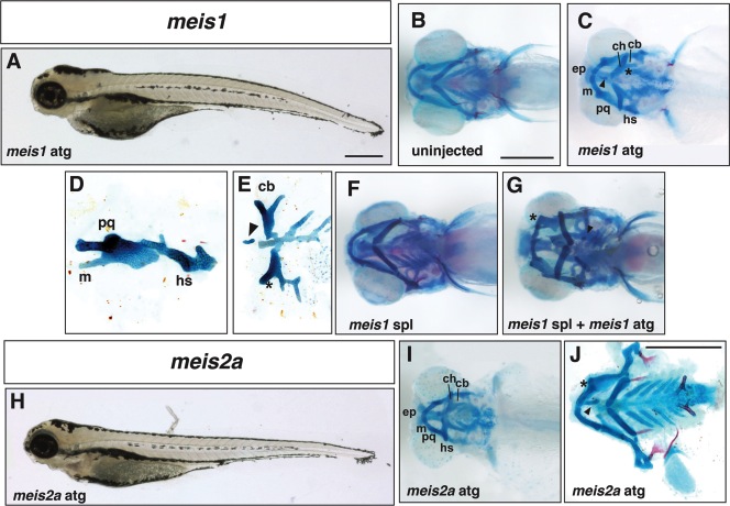
Fig. 3 Defects in cranial development in Class I meis morphants. A: Lateral view of a 5dpf larva injected with 5 ng meis1 ATG Morpholino. B: Ventral view of the craniofacial skeleton of an uninjected larva at 5dpf. C: Ventral view of a meis1 ATG morphant showing a fusion between the Meckel′s and palatoquadrate cartilages and the ceratohyal and first ceratobranchial (asterisk). Ectopic cartilage is indicated by an arrowhead. D: Lateral view of a flat mount showing a fused Meckel′s and palatoquadrate cartilage and the hyosymplectic. E: Ventral view of flat mount showing ectopic cartilage (arrowhead) and fused ceratobranchials (asterisk). F: Ventral view of a larval skeleton 5 days after injection with 20 ng meis1 SPL Morpholino. G: Ventral view of a larval skeleton after co-injection with 20 ng meis1 SPL Morpholino and 2 ng meis1 ATG Morpholino. Ectopic cartilage is indicated by an arrowhead and the fusion between Meckel′s and the palatoquadrate is indicated by the asterisk. H: Lateral view of a 5dpf larva injected with 10 ng meis2a Morpholino. I: Ventral view of the skeleton of a meis2a ATG morphant. J: Flat mount of a meis2a morphant (5 ng ATG MO) indicating a fusion between Meckel′s and palatoquadrate (asterisk) and ectopic cartilage (arrowhead). Abbreviations as in Figure 1.