
|
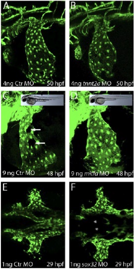
Fig. S5 CCV development is not affected by blood flow, melanocytes or endoderm. (A-F) Confocal fluorescent images of transgenic zebrafish embryos at the indicated time points. ECs were visualized by transgenic EGFP expression [Tg(kdrl:EGFP)s843]. (A-D) Lateral views; (E,F) dorsal views. (A,B) Embryos injected with control MO (A) or with tnnt2a MO (B). Note that the CCV developed normally in the absence of blood flow in tnnt2aMO-injected embryos (B). (C,D) Embryos injected with control MO (C) or mitfa MO (D). Note that although mitfa MO-injected embryos had no melanocytes (compare brightfield images, insets in C and D) the CCVs developed normally. Arrows indicate melanocytes in C. (E,F) Embryos injected with control MO (E) or with sox32 MO (F). Note that in the absence of endoderm the LDA fail to form (asterisks), whereas the CCVs develop (F). CCV, common cardinal vein; Ctr, control; LDA, lateral dorsal aortae; WT, wild type.