
|
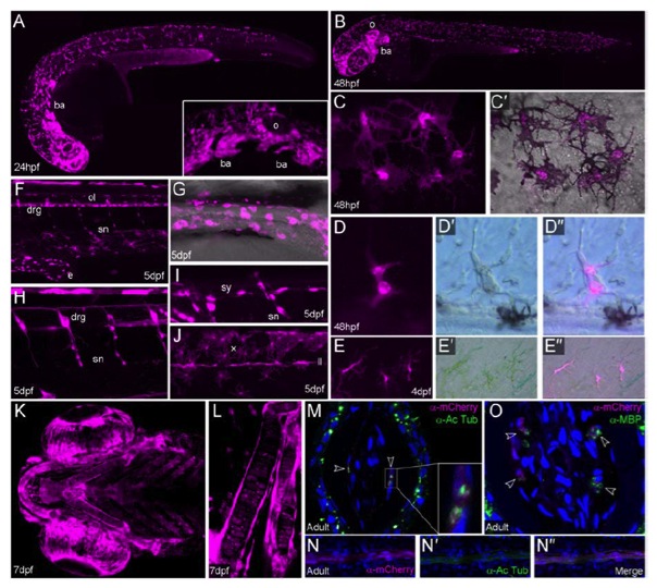
Fig. S1 The sox10:cre; ubi:switch line permanently labels all neural crest derivatives
A-L: Images of sox10:cre; ubi:switch embryos and larvae at 24hpf (A), 48hpf (B-D′′), 4dpf (E-E′′), 5dpf (F-J) and 7dpf (K-L). Live confocal (A-C′, F-L) and epifluorescent (D-E′′) images showing 2 mCherry expression in magenta. All are viewed laterally, except K and L which are ventral views. Brightfield views (D′, E′′) and overlays (D′′,E′′) are shown to visualise pigmentation. Neural crest cells can be first observed expressing mCherry weakly from 24hpf (A) and mCherry levels accumulate from there onwards, such that by 48hpf there is comprehensive expression in migrating neural crest cells (B). Neural crest cells in the branchial arches (ba) are labelled (A(inset), B). This line additionally labels the sox10 expressing otic vesicle (o; A(inset)-B). All three chromatophore lineages are broadly labelled, namely black melanophores (C-C′), iridescent iridophores (D-D3) and yellow xanthophores (E- E′′, J). As melanin strongly quenches the fluorescent signal, we visualised melanophores in young or partially PTU treated embryos. Counts of melanophores in 5 larvae at 54hpf showed 96.6 ± 0.4% were visibly mCherry positive (506 cells counted). There is also extensive labelling of the neural crest derived peripheral nervous system, comprising the enteric nervous system (e; F, G), peripheral glia of both the spinal nerves (sn; F, H, I) and the lateral line (ll; J), sympathetic ganglia (sy; I), and dorsal root ganglia (drg; F, H). There is widespread expression in the cells of the cranial skeletal elements (K, L). Ventral view of the pharyngeal elements generated by projection of confocal stacks, showing mCherry is found in all elements of the visceral cranium. Magnified view demonstrates expression is seen in both central stacks of cartilage cells and lining osteoblasts (L). M-O: Images of the fin rays of sox10:cre; ubi:switch adults co-immunostained for mCherry (magenta) and either Acetylated tubulin (M-N3 green) or Myelin Basic Protein (O; green). Lateral micrographs (N-N′′) and transverse cryosections (M) demonstrate close association of neural crest derived mCherry cells with axons within the inner surface of the fin rays. The mCherry cells appear to surround the axons (M – arrowheads and inset). Co-labelling with Myelin Basic Protein (MBP; O - arrowheads) supports the identity of these mCherry cells as Schwann cells.