
|
Fig. S5
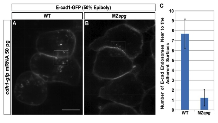
Number of E-cad Endosomes Adjacent to Adherent Surfaces is smaller in MZspg Blastoderm Cells than WT
(Related to Figure 7)
In vivo immunofluorescence and quantification of E-cad1-GFP endosomal vesicles near to adherent surfaces between deep cells in WT and MZspg cells. WT (A) and MZspg (B) embryos were labeled by injection of cdh1-gfp mRNA into one cell at 8-cell stage. The scatter labeled embryos developed until 50% epiboly stage, and E-cad1-GFP fluorescence was documented in vivo by confocal microscopy. On the images, fluorescent spots were automatically detected using Imaris software. Spots adjacent to adherent surfaces (boxes) were counted as endosomes (C). In MZspg cells, most spots were located directed in the adherent surface, which were not counted as endosomes (B). The average number of E-cad endosomes adjacent to the adherent surfaces was significantly smaller in MZspg compared to WT embryos (C) (P < 0.001; n = 61 focal planes each in 12 WT and 11 MZspg embryos). Animal views. Error bars represent standard error of the mean (SEM). Scale bar = 10 μm.
Reprinted from Developmental Cell, 24(5), Song, S., Eckerle, S., Onichtchouk, D., Marrs, J.A., Nitschke, R., and Driever, W., Pou5f1-dependent EGF expression controls e-cadherin endocytosis, cell adhesion, and zebrafish epiboly movements, 486-501, Copyright (2013) with permission from Elsevier. Full text @ Dev. Cell