
Image
|
Figure Caption
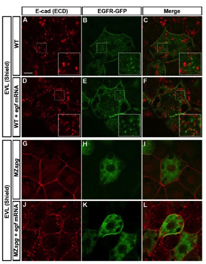
Fig. S4
Comparison of EGF-mediated Vesicular Trafficking of E-cad and EGFR-GFP
(Related to Figure 5)
(A-L) Co-immunofluorescence of E-cad (anti ECD; red) and EGFR-GFP (green) in WT (A-C), egf mRNA injected WT (D-F), MZspg (G-I), and egf mRNA injected MZspg (J-L) embryos at shield stage in fixed whole mounts. Merged channels reveal no colocalization between E-cad and EGFR positive endosomes in WT and MZspg embryos, as well as in WT and MZspg embryos overexpressing EGF. Insets in A-F show higher magnification. Animal pole views. Scale bar = 10 μm.
Acknowledgments
This image is the copyrighted work of the attributed author or publisher, and
ZFIN has permission only to display this image to its users.
Additional permissions should be obtained from the applicable author or publisher of the image.
Reprinted from Developmental Cell, 24(5), Song, S., Eckerle, S., Onichtchouk, D., Marrs, J.A., Nitschke, R., and Driever, W., Pou5f1-dependent EGF expression controls e-cadherin endocytosis, cell adhesion, and zebrafish epiboly movements, 486-501, Copyright (2013) with permission from Elsevier. Full text @ Dev. Cell