
Image
|
Figure Caption
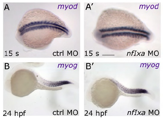
Fig. S2 First myogenic wave takes place correctly in nfixa-MO-injected embryos. (A-B′) myod (A,A′) and myog (B,B′) expression patterns in nfixa-MO embryos during somitogenesis and at 24 hpf are the same as control, suggesting that the segmentation process and the first myogenic wave are correctly formed. In A,A′, dorsal views are shown; in B,B′ lateral views are shown. Anterior is always towards the left. Scale bars: 100 μm in A,A′; 150 μm in B,B′.
Figure Data
Acknowledgments
This image is the copyrighted work of the attributed author or publisher, and
ZFIN has permission only to display this image to its users.
Additional permissions should be obtained from the applicable author or publisher of the image.
Full text @ Development