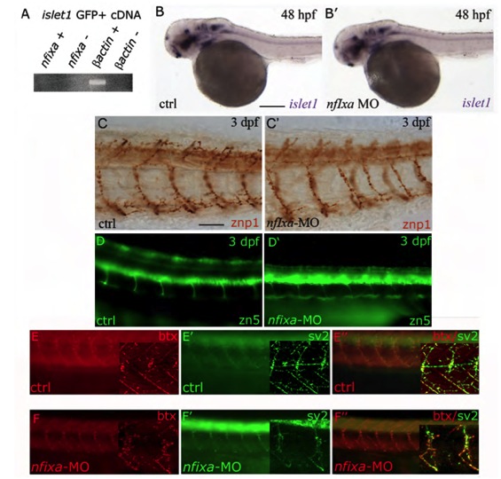
Fig. S3 Nfixa loss of function does not influence motoneurons development (A) RT-PCR performed on cDNA of motoneurons sorted from islet1 GFP transgenic embryos at 48 hpf. nfixa is not expressed in this cell population. (B,B′) islet1 expression pattern in nfixa-MO embryos at 48 hpf does not present differences in comparison with control embryos, suggesting that motoneurons are correctly formed. (C-D′) Axonal projections of primary (C,C′) and secondary motoneurons (D,D′) visualized by znp1 and zn-5 antibodies, respectively, are correctly formed in nfixa-MO-injected embryos. (E-F′′) Synapses were labeled with bungarotoxin (BTX red, postsynaptic AChRs) and SV2 (green, presynaptic vesicles) antibodies. The co-localization of both signals in control (E′′) and nfixa-MO injected (F′) embryos showed functional neuromuscular synapses. Main panels are images taken on a fluorescence microscope; insets show single-plane confocal images/projections from a confocal z-stack.
Image
Figure Caption
Figure Data
Acknowledgments
This image is the copyrighted work of the attributed author or publisher, and
ZFIN has permission only to display this image to its users.
Additional permissions should be obtained from the applicable author or publisher of the image.
Full text @ Development

