
Image
|
Figure Caption
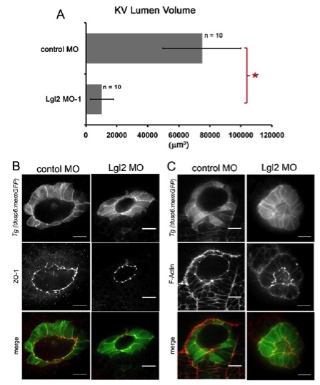
Fig. S2 Depletion of Lgl2 reduced KV lumen volume, but did not alter localization of apical epithelial markers. (A) Analysis of KV lumen volume at the 8-somite stage in live Tg(dusp6:memGFP) embryos. Error bars represent s.d. n, number of embryos analyzed. *P<0.05. (B,C) Fluorescence staining of apical markers in Tg(dusp6:memGFP) embryos injected with control MO or Lgl2 MO. ZO-1 (B) and F-Actin (C) were localized at apical membranes lining the KV lumen in both control and Lgl2-depleted embryos. Scale bars: 20 μm.
Acknowledgments
This image is the copyrighted work of the attributed author or publisher, and
ZFIN has permission only to display this image to its users.
Additional permissions should be obtained from the applicable author or publisher of the image.
Full text @ Development