
|
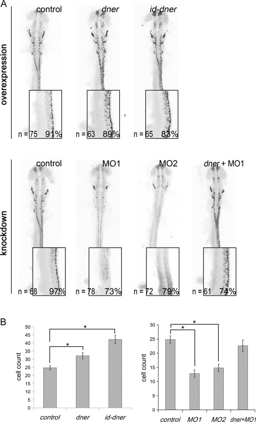
Fig. 4 Dner-induced neuronal differentiation and dner morpholinos are sufficient to inhibit differentiation. Embryos were analyzed by immunohistochemistry using anti-Hu antibody at 24 hpf. The black-and-white fluorescent signals were inverted using negative film for a better presentation. (A) Injections with either dner or id-dner increased the frequency of HuC/D-positive cells. Injections with either morpholino were sufficient to decrease the HuC/D signals. This reduction could be rescued by co-injection with dner cRNA. The insets in each panels show lateral view enlargements of the 3-somite to 9-somite levels of the spinal cord. HuC/D-positive cells were counted in this region and quantified in (B). N, p<0.05.
Reprinted from Developmental Biology, 375(1), Hsieh, F.Y., Ma, T.L., Shih, H.Y., Lin, S.J., Huang, C.W., Wang, H.Y., and Cheng, Y.C., Dner inhibits neural progenitor proliferation and induces neuronal and glial differentiation in zebrafish, 1-12, Copyright (2013) with permission from Elsevier. Full text @ Dev. Biol.