
|
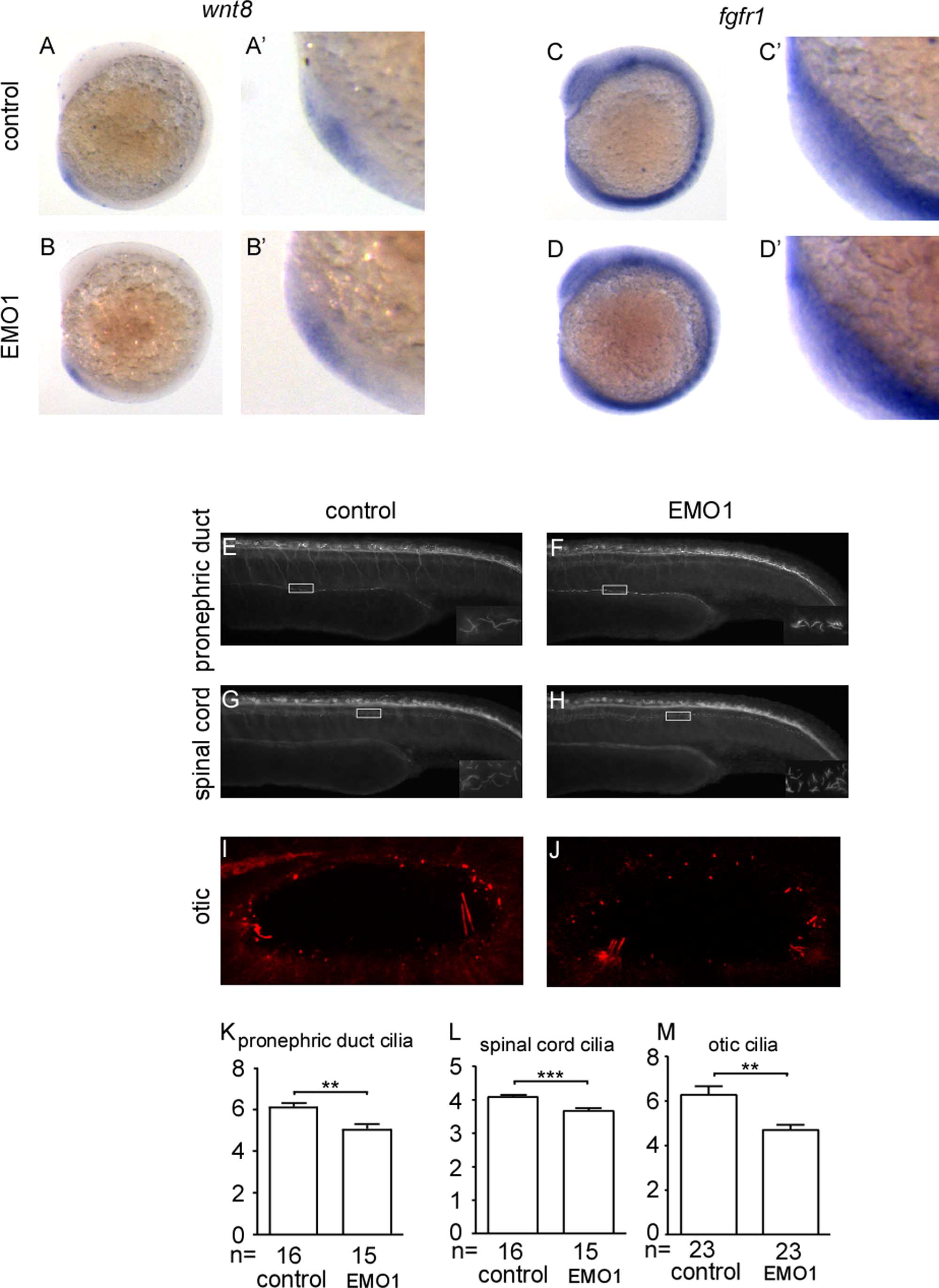
Fig. S3 Knockdown of enc1l does not affect the midline, KV structure formation or KV cell number. (A): There is no difference in the KV marker sox32 between enc1l-MO injected embryos and control embryos. (B) No difference in the midline marker ntl is detected between enc1l morphants and control embryos. Injected enc1l-MO2 (D) can decrease the cilia length and increase the cilia number compared to control (C) embryos. (E): Statistical graph of the cilia number. (F): Statistical graph of the cilia length. Enc1l-MO1 (G) and control (H) embryos stained with ace-tubulin (green), phalloidine (red), and DAPI (blue). (I): Statistical graph of the KV cell number. ***: p<0.005.
Reprinted from Developmental Biology, 374(1), Qian, M., Yao, S., Jing, L., He, J., Xiao, C., Zhang, T., Meng, W., Zhu, H., Xu, H., and Mo, X., ENC1-like Integrates the Retinoic Acid/FGF Signaling Pathways to Modulate Ciliogenesis of Kupffer's Vesicle during Zebrafish Embryonic Development, 85-95, Copyright (2013) with permission from Elsevier. Full text @ Dev. Biol.