
|
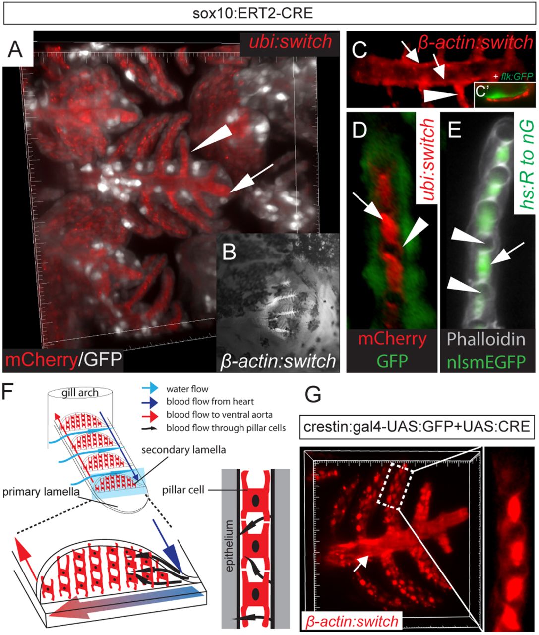
Fig. 5 NC origin of gill pillar cells. (A-E) Recombined cells in the primary and secondary lamellae of the gills. (A) 3D view of confocal stacks showing the gill lamellae in an induced Tg(sox10:ERT2-Cre;ubi:switch) adult fish: non-NC-derived tissues are in gray (GFP), whereas NC-derived tissues in the primary (arrow) and in the secondary (arrowhead) lamellae are in red (mCherry). (B) Epifluorescence image of DsRed+ cells in the primary and secondary lamellae in an induced Tg(sox10:ERT2-Cre;β-actin:switch) adult fish. (C) Confocal section of labeled smooth muscles of the blood vessel tunica media in the primary lamellae (arrows) and pillar cell in contact with the smooth muscle layer (arrowhead). (C2) flk:GFP+ endothelium in the gills surrounded by NC-derived smooth muscles. (D) Confocal section of a secondary lamellae showing recombined mCherry+ pillar cells surrounded by non-recombined GFP+ epithelial cells in an induced Tg(sox10:ERT2-Cre;ubi:switch) adult fish. The arrow indicates the pillar cell body, whereas the arrowhead marks a cytoplasmic process connecting two neighboring pillar cells. (E) MIP of confocal stacks showing a phalloidin-stained (gray) secondary lamella with the GFP+ pillar cell nuclei of an induced hs:R to nG adult fish. The pillar cell nuclei (arrow) are located between adjacent lumina (arrowheads). (F) Schematic of gill lamellae and pillar cells demonstrating the flow of blood through the organ. (G) MIP of confocal stacks of recombined lamellae using the crestin promoter, showing labeled smooth muscles (arrow) and pillar cells (inset).