
|
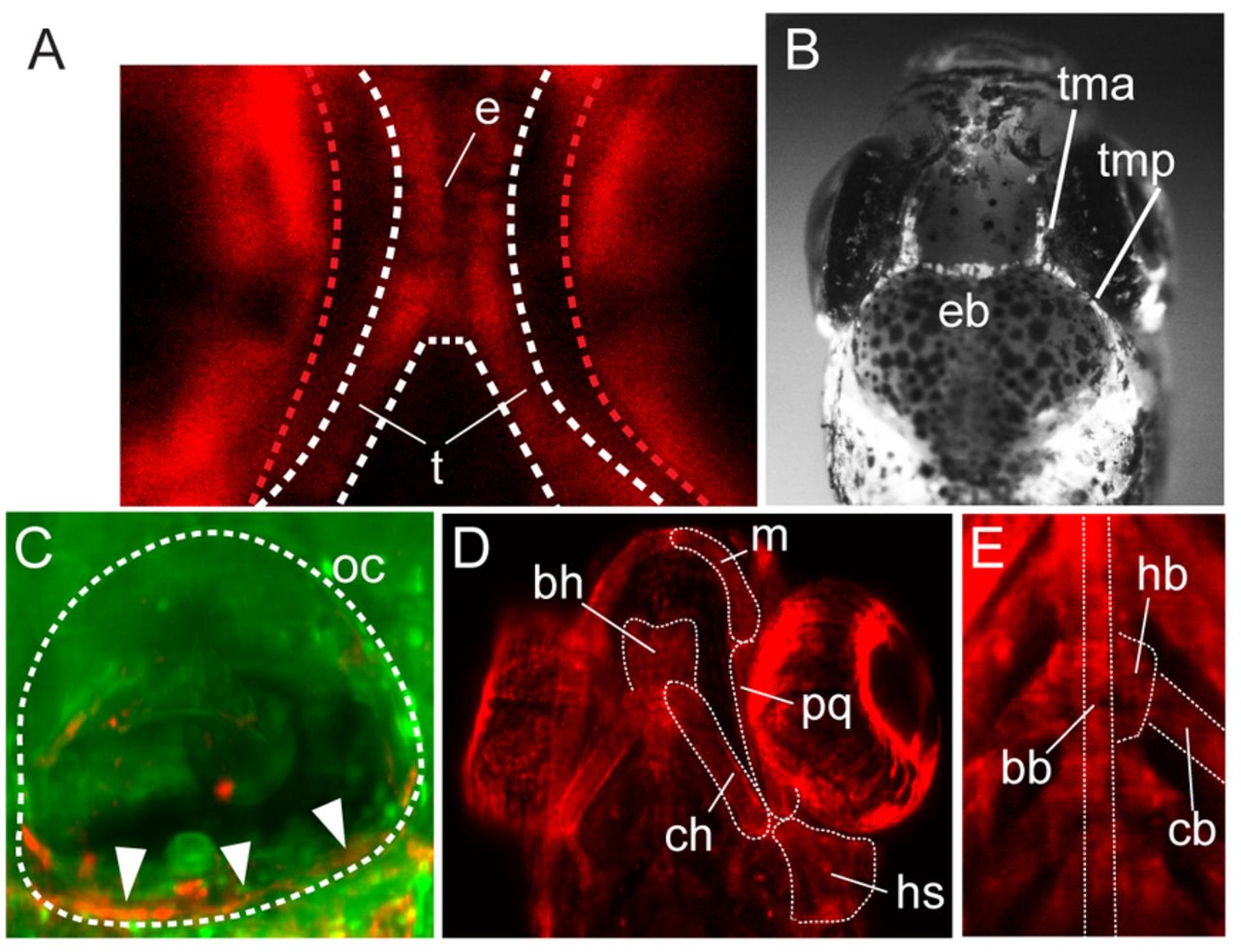
Fig. 3 NC-derived elements in the developing chondrocranium and viscerocranium. (A) Confocal section of recombined tissue in the ethmoid plate (e) and the trabeculae (t) of a 5-dpf Tg(sox10:ERT2-Cre;ubi:switch) larva. The red dashed lines highlight the autofluorescence of the eyes. (B) Epifluorescent image of labeled elements of the dorsal neurocranium in 15-dpf Tg(sox10:ERT2-Cre;β-actin:switch) fish: taenia marginalis anterior (tma), taenia marginalis posterior (tmp) and epiphyseal bar (eb). (C-E) MIP of confocal stacks. (C) Recombined cells in the forming ventral cartilage of the otic capsule (oc, arrowheads) in 5-dpf Tg(sox10:ERT2-Cre;ubi:switch) larvae. (D) Labeled viscerocranial elements in the mandibular region [Meckel′s cartilage (m), palatoquadrate (pq)] and in the hyod region [hyosymplectic (hs), ceratohyal (ch), basihyal (bh)] of 5-dpf Tg(sox10:ERT2-Cre;ubi:switch) larvae. (E) Labeled basibranchial (bb), hypobranchial (hb) and ceratobranchial (cb) elements of 5-dpf Tg(sox10:ERT2-Cre;ubi:switch) larvae.