
Image
|
Figure Caption
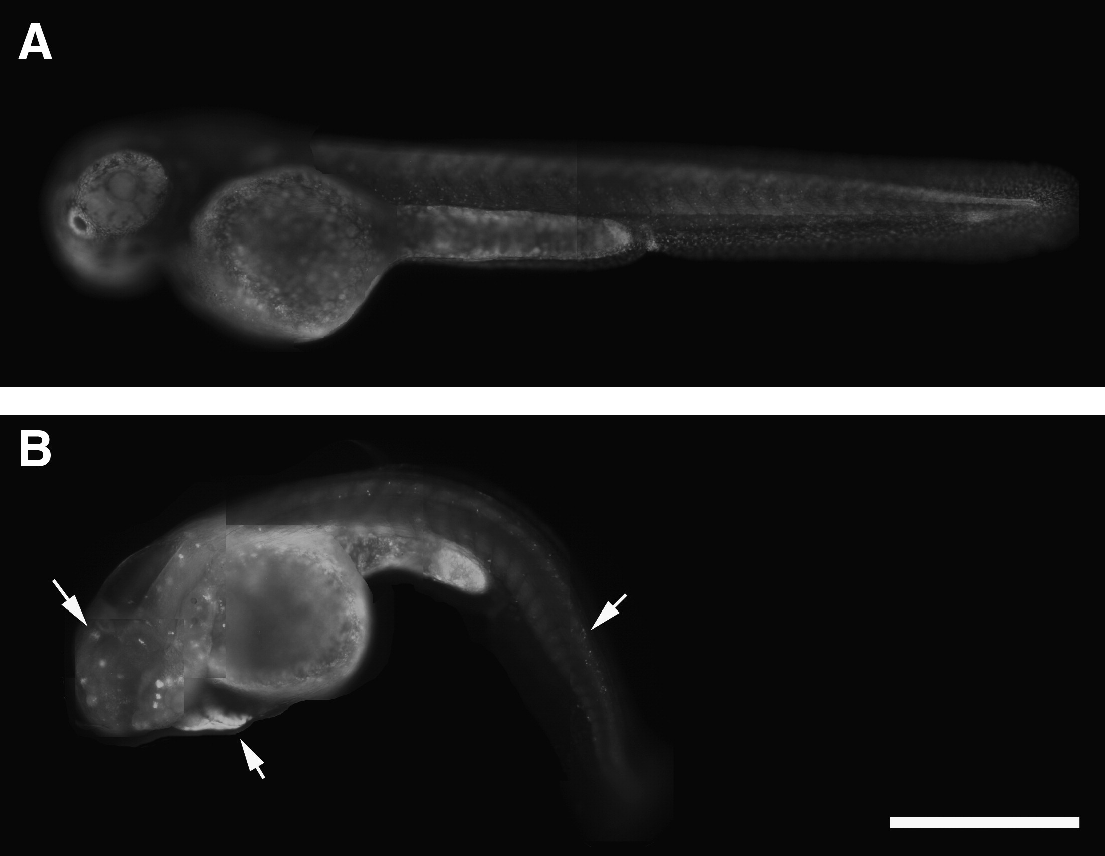
Fig. 8 Carbaryl-treated embryos experience an increase in cell death. Lateral views of MeOH control (A) and carbaryl-treated (B) embryos exposed to acridine orange at 48 hpf. Dying cells near the heart, in the head region, and along the spinal cord are apparent in carbaryl-treated embryos (B; arrows), and far exceed the amount of dying cells observed in MeOH control embryos. Different focal plane images were merged in Photoshop to produce complied images. Scale bar: 500 μm.
Acknowledgments
This image is the copyrighted work of the attributed author or publisher, and
ZFIN has permission only to display this image to its users.
Additional permissions should be obtained from the applicable author or publisher of the image.
Full text @ Zebrafish