
|
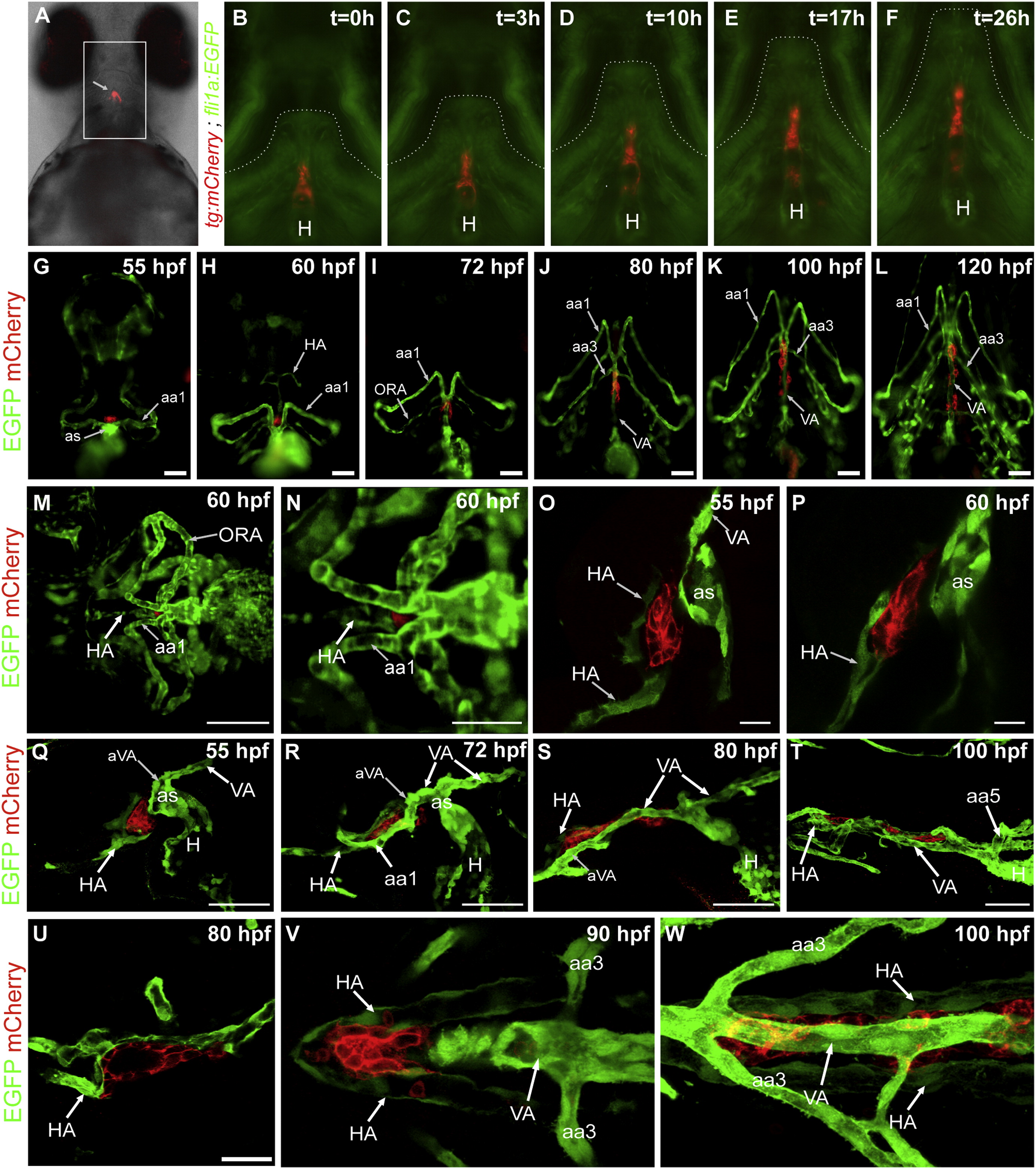
Fig. 4 Rostral expansion of thyroid tissue along the pharyngeal midline occurs coordinatedly with pharyngeal vessel remodeling. (A) Ventral view of a live tg(tg:mCherry) embryo at 70 hpf displaying strong red fluorescence of the rostrally expanding thyroid tissue (arrow). ((B)-(F)) Stills from a time-lapse movie of a tg(tg:mCherry;fli1a:EGFP) embryo showing that thyroid expansion along the pharyngeal midline occurs concomitantly with a general remodeling of the hypobranchial region. EGFP labels endothelial cells and neural crest-derived mesenchyme. Live imaging was started at 75 hpf and lasted for 26 h. The timer indicates the time elapsed since the beginning of live imaging. Ventral views are shown, anterior is to the top. ((G)-(L)) Whole-mount IF staining of GFP and mCherry for a series of fixed tg(tg:mCherry;kdrl:EGFP) embryos shows developmental changes in gross thyroid morphology relative to the remodeling pharyngeal vasculatures. EGFP labels endocardium and vascular endothelium. Ventral views are shown and anterior is to the top. ((M) and (N)) Live imaging of tg(tg:mCherry;kdrl:EGFP) embryos by selective plane illumination microscopy (SPIM) identified the hypobranchial artery (HA) as the main vessel contacting the thyroid during the initial stages of its rostral expansion. (M) shows a single frame from time-lapse Movie 6. (N) shows a magnified view of the outflow tract (OFT) region. M and N show ventral views, anterior is to the left. ((O) and (P)) The close contact between thyroid cells and endothelial cells of the forming HA was confirmed by confocal microscopy of tg(tg:mCherry;kdrl:EGFP) embryos after IF staining for mCherry and GFP. Confocal sections of sagittal vibratome sections are shown, anterior is to the left. ((Q)–(W)) Confocal microscopy of a developmental series of tg(tg:mCherry;kdrl:EGFP) embryos after IF staining for mCherry and GFP demonstrate the location of thyroid tissue relative to the HA and VA. During the initial rostral expansion of the thyroid (55–70 hpf), the thyroid was in close contact with the HA while the VA, along its entire length, was still located caudal to the heart OFT. Thereafter, a progressive rostral protrusion of the VA relative to the OFT was evident (see (R)–(T)). Between 70 and 75 hpf, the anterior end of the VA (aVA) was located at the level of the thyroid, while the major part of the VA was still located caudal to the OFT (see (R)). Around 100 hpf, the entire VA was located anterior to the OFT (see (T)). By that time, the rostral limit of the VA was located anteriorly to the thyroid. Note that the location of the most anterior thyroid tissue was invariably determined by the location of the HA branching point (see (O),(P),(U) and (V)). The caudally running paired HA demarcates the lateral extension of the dispersed thyroid follicles (see (W)). Panels (Q)-(U) show confocal images acquired from sagittal vibratome sections. Panels (V) and (W) show confocal images acquired from frontal vibratome sections. aa1, aa3, aa5, aortic arch arteries 1, 3, 5; as, aortic sac; (H), heart; HA, hypobranchial artery; ORA, opercular artery; VA, ventral aorta. Scale bars: 50 μM in panels (G)-(L), (N), (Q)–(W), 100 µM in panel M, 20 μM in panels (O) and (P).
Reprinted from Developmental Biology, 372(2), Opitz, R., Maquet, E., Huisken, J., Antonica, F., Trubiroha, A., Pottier, G., Janssens, V., and Costagliola, S., Transgenic zebrafish illuminate the dynamics of thyroid morphogenesis and its relationship to cardiovascular development, 203-216, Copyright (2012) with permission from Elsevier. Full text @ Dev. Biol.