
|
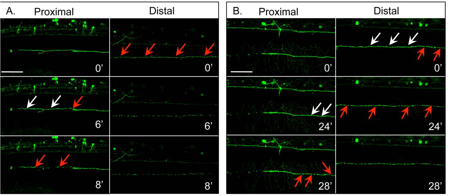
Fig. S3 Fragmentation proceeds in a distal-to-proximal direction. Single axons of fish injected with HuC::GFP were transected at 72 hpf, and imaged every 2 minutes both adjacent to the axotomy site (proximal) and further down the length of the embryo (distal). A and B are images from two representative fish mounted laterally, with their left side visible. Fragmentation was observed (red arrows) in the distal segment earlier than in the proximal segment. Beading (white arrows), which precedes fragmentation, was also seen first in the distal segment. The length of time between the onset of fragmentation in distal and proximal segments is variable. Times shown are relative to the first image in each set. Scale bar, 100 μm.