
Image
|
Figure Caption
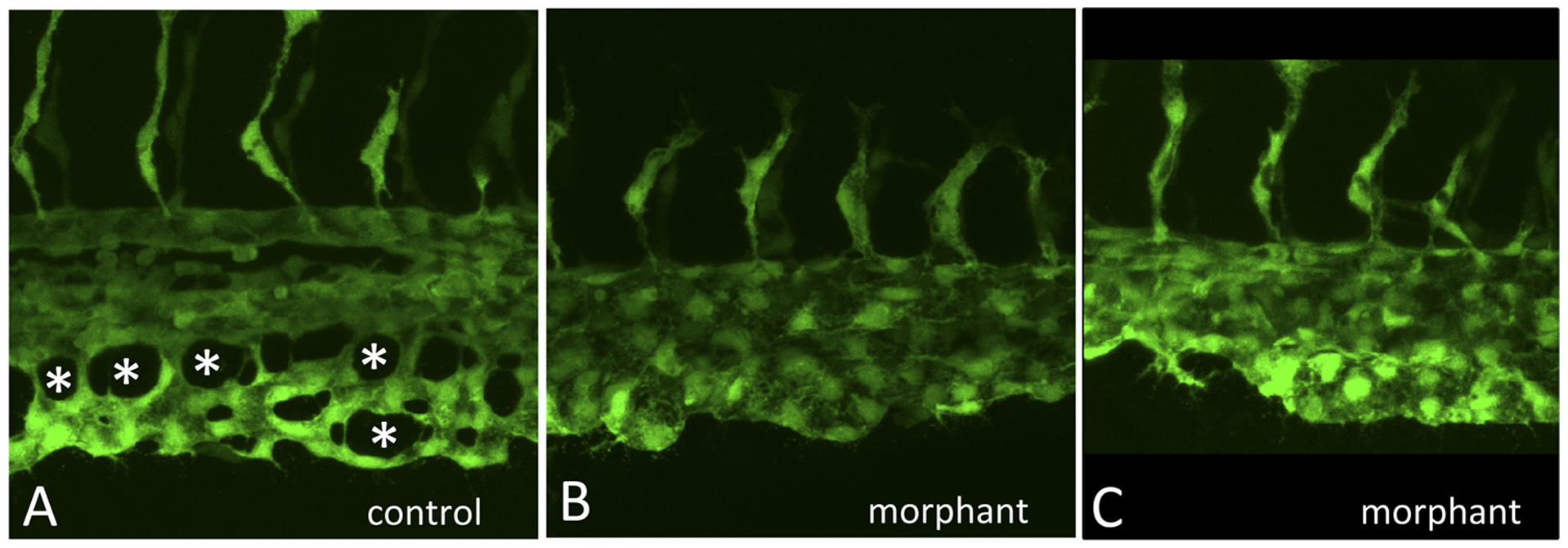
Fig. 3 The gata4 morphant CHT fails to develop fenestrated vascular structures.
Shown are higher resolution images of control (A) and two independent examples of morphant embryos (B,C) imaged at 32 hpf by confocal microscopy, as the caudal plexus is forming. While vasculogenesis initiates normally, and intersegmental vessels sprout equivalently, angiogenesis in the CHT of control embryos generates a vascular plexus containing open fenestrated structures, indicated by the asterisks in A, that are missing in gata4 morphants.
Figure Data
Acknowledgments
This image is the copyrighted work of the attributed author or publisher, and
ZFIN has permission only to display this image to its users.
Additional permissions should be obtained from the applicable author or publisher of the image.
Full text @ PLoS One