
|
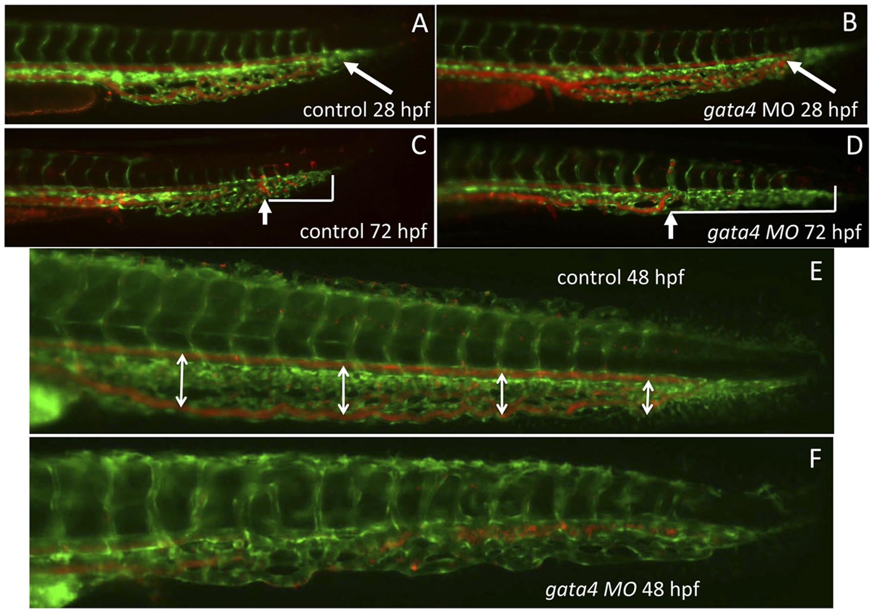
Fig. 2 The vascular morphology of the CHT is disrupted in the gata4 morphant embryo.
Shown are representative embryos (n >50) derived from tg(gata1:dsred; fli1:egfp) double transgenic reporter fish. The green signal shows the vascular endothelium, while the red signal shows the circulation of blood cells, which are moving so quickly that individual cells are not seen. At 28 hpf (A,B) the blood circulates to the caudal end of the developing CHT (the caudal end is indicated by white arrows) in both control uninjected (A) and gata4 morphant (B) embryos. However, by 72 hpf (C,D), the vascular plexus is much more developed in the control (C) compared to the morphant (D). In addition, the blood circulation is restricted in the morphant, failing to move into the more caudal regions, as indicated in panels C and D by the white brackets that mark the distance from the caudal end of the CHT to the most caudal position of circulating cells (indicated by the white arrows). The block to plexus formation can be seen clearly at 48 hpf (E,F), when in the control embryos (E) the dorsal aorta and caudal vein are well separated to create a fenestrated space (this space is indicated by double-headed arrows), which is not seen in the morphant (F). MO = morphant. Views are lateral, anterior to the left.