
|
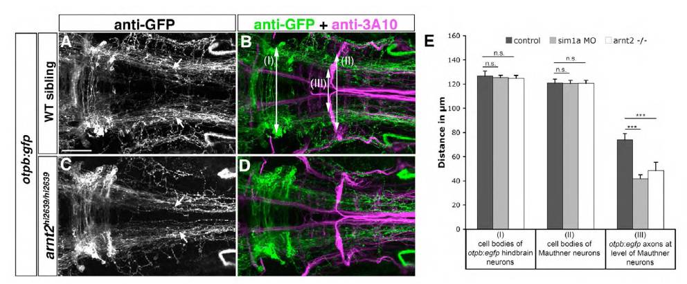
Fig. S2 Lateral positioning of longitudinal otpb:gfp-positive axons is altered in arnt2hi2639c mutants. Dorsal views of confocal z projections of the hindbrain of otpb:gfp transgenic embryos co-labeled with anti-GFP and anti-3A10 (A-D) at 72 hpf are shown. (A,B) Wild-type siblings display normal medio-lateral positioning of otpb:gfp positive axons (arrows in A). (C,D) In arnt2hi2639c/hi2639c homozygous mutants, longitudinal projections of otpb:gfp axons are shifted towards the midline (arrows in C). Midline crossing of Mauthner axons is not affected in arnt2hi2639c mutants, suggesting grossly normal hindbrain development (compare arrowheads in B,D). (E) Quantification of mediolateral positioning of otpb:gfppositive longitudinal axons at the anterior-posterior level of Mauthner neurons (see III in B), of the distance of MA neurons (see II in B) and of otpb:gfp hindbrain neurons (see I in B) in arnt2hi2639c/hi2639c homozygous mutants or wild-type siblings. Numbers in parentheses indicate the number of embryos analyzed. ***P<0.0001; n.s., not significant. Scale bar: 50 μm.