
|
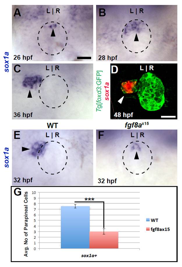
Fig. S2 sox1a is expressed in parapineal cells near their time of differentiation. All images are from dorsal view. (A) sox1a expression (black arrowhead) in WT pineal complex anlage (dashed circle). (B) A cluster of sox1a-expressing cells (black arrowhead) are in the anterior pineal complex anlage of 28 hpf WT embryos. (C) By 36 hpf, sox1a expression is present (black arrowhead) to the left of the pineal anlage suggesting that it is indeed expressed in parapineal cells. (D) Confocal slice of 48 hpf WT larvae showing colocalization of sox1a (white arrowhead) and Tg[foxd3:GFP] confirming that sox1a is expressed in parapineal cells. (E,F) sox1a expression (black arrowheads) in WT and fgf8ax15 mutants at 32 hpf. (G) Graph quantifying the number of sox1a-expressing cells in WT and fgf8ax15 mutants at 32 hpf. fgf8ax15 mutants have significantly reduced numbers of sox1a-expressing cells (***P<0.0005 by t-test). Error bars represent s.e.m. Scale bar: in A, 20 μm; in B, 25 μm; in C: 20 μm.