
|
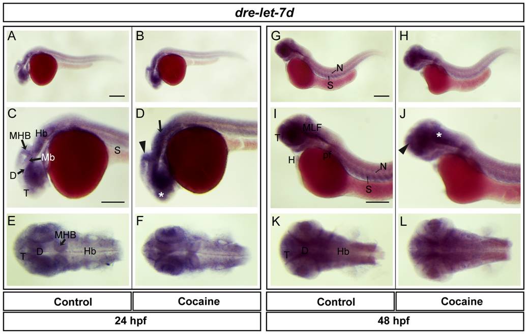
Fig. 10 Spatial distribution of dre-let-7d at 24 and 48 hpf.
Lateral views (A-D, G-J), dorsal views (E-F, K-L). dre-let-7d was found in the telencephalon, diencephalon, midbrain (optic tectum), MHB, hindbrain, spinal cord, eye and somites. Expression in embryos exposed to cocaine was observed in the telencephalon, MHB, and hindbrain (asterisk, arrowhead and arrow, respectively). At 48 hpf, dre-let-7d was expressed to a similar extent to the situation at 24 hpf, but also with expression in the MLF, notochord, pectoral flipper and heart. Cocaine induced an increase in expression in the telencephalon and MLF (arrowhead and asterisk, respectively). Abbreviations: T: telencephalon; Mb: midbrain, Hb: hindbrain, D: diencephalon, N: notochord; S: somite; pf: pectoral flipper; H: heart. Scale bar: 200 μm in A (Applies to B, G and H) and 250 μm in C (Applies to D, E, F, I, J, K and L).