
|
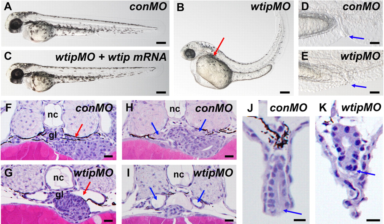
Fig. 2 wtip knockdown embryos show pronephric cyst formation accompanied by cloaca malformation, hydrocephalus, body axis curvature and pericardial edema.
(A,B,C) Side view of embryos at 48 hpf under light microscopy. (B) 48 hpf wtip morphants form pronephric cysts, hydrocephaly, body curvature and pericardial edema. The red arrow marks the location of the cyst dilation (B). (D,E) Lateral view of cloaca at 48 hpf. The blue arrow marks the cloaca. (C) wtip mRNA can rescue pronephric cyst, body curvature, hydrocephalus, and pericardial edema caused by wtipMO. Histological transverse-sections of 48 hpf embryos are 4µm JB-4 plastic sections stained with hematoxylin and eosin (F–K) at the level of the glomerulus (F,G; red arrow), anterior pronephros (H,I; blue arrow), and cloaca (J,K; blue arrow). Glomerular cysts (G), dilated anterior pronephros (I) and cloaca malformation (K) were observed in the 48 hpf wtip morphants. Control morpholino injected embryos (conMO), wtip morpholino injected embryos (wtipMO), glomerulus (gl), and notochord (nc). Scale bars are 200 μm in A–C, 500 μm in D,E, and 100 μ in F–K.