
|
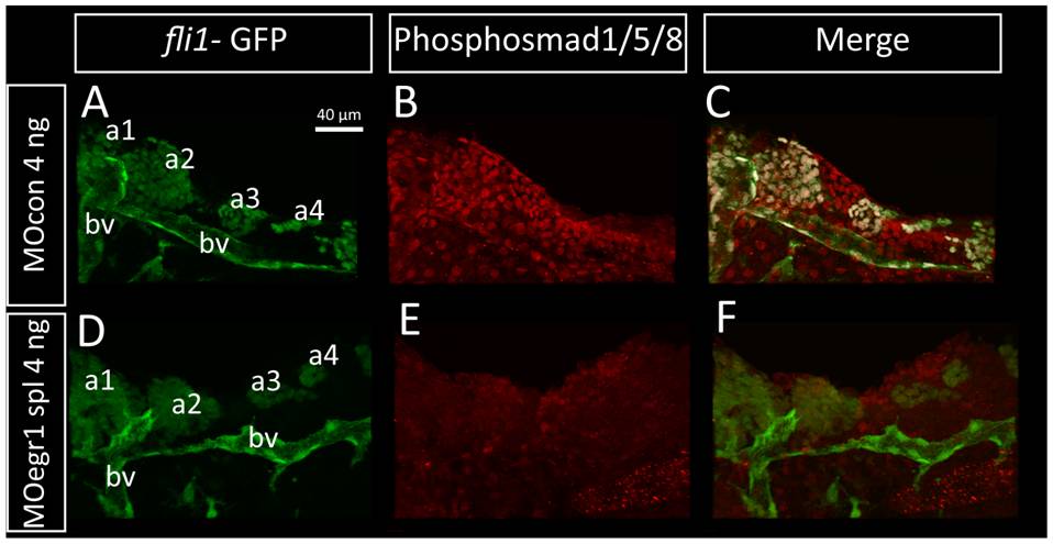
Fig. 10 Bmp signaling is down-regulated in egr1 morphants.
Pharyngeal cartilage precursor cells were visualized by immunohistochemistry using anti-GFP antibodies (green) in fli-GFP embryos. Activity of the BMP signaling pathway was assessed using antibodies against phospho-Smad1/5/8 (red) in 32 hpf embryos. Ventral view of pharyngeal arches, scale bar 40 μm. (A-F) Pharyngeal cartilage precursor cells were visualized by immunohistochemistry using anti-GFP antibodies (green) in fli1-GFP embryos. Activity of the BMP signaling pathway was assessed using antibodies against phospho-Smad1/5/8 (red) in 32 hpf embryos. Ventral view of pharyngeal arches, scale bar 40 μm. (A,B,C) 4 ng MOcon injected embryos, (D, E, F) 4 ng MOegr1 spl injected embryos. fli1-GFP embryos express the GFP transgene in cartilage precursors and endothelial cells in control (A) and in egr1 morphants (D). In contrast, phospho-Smad1/5/8 is is clearly down regulated in egr1 morphants (E) compared to control embryos (B). (C,F) Overlay images of the two anti-body signals clearly show that phospho-Smad1/5/8 is present in GFP-epressing cartilage precursor cells in control embryos (C), while no colocalization is observed in egr1 morphants (F). (a1) first arch, (a2) second arch, (a3) third arch, (a4) fourth arch, (bv) blood vessel.