
Image
|
Figure Caption
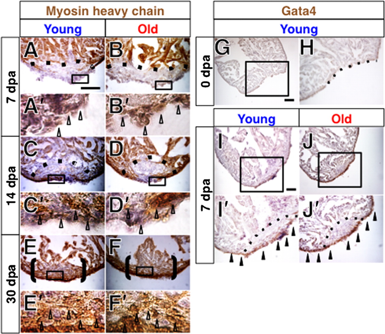
Fig. 2 Regeneration of the myocardial layer and induction of Gata4 expression in young and old fish hearts.
(A-F′) MHC staining of regenerating hearts at 7 dpa (A–B′), 14 dpa (C–D′) and 30 dpa (E-F′) in young (A,A′,C,C′,E,E′) and old (B,B′,D,D′,F,F′) fish. A′-F′ show higher magnification images of the boxed areas in A-F. Open arrowheads indicate MHC signals in the regenerating area. For simplicity, not all signals are pointed. Scale bar: 50 μm; the degree of zoom: ×5.5. (G,H) Gata4 immunoreactivity was not detected immediately after amputation. H shows a close up of the boxed areas in G. Scale bar: 50 μm; the degree of zoom: ×2. (I-J′) Gata4 staining of regenerating hearts at 7 dpa in young (I,I′) and old (J,J′) fish. Black arrowheads indicate Gata4 signals. I′ and J′ show close ups of the boxed areas in I and J. Sections were counterstained with hematoxylin. Dotted lines indicate the amputated planes of 7 and 14 dpa hearts. Brackets indicate the regenerating areas of 30 dpa hearts. Scale bar: 50μm; the degree of zoom: ×2.
Acknowledgments
This image is the copyrighted work of the attributed author or publisher, and
ZFIN has permission only to display this image to its users.
Additional permissions should be obtained from the applicable author or publisher of the image.
Full text @ Biol. Open