
Image
|
Figure Caption
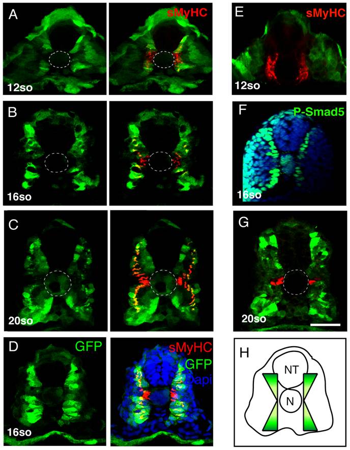
Fig. 6 BMP signaling forms a dorsal and ventral gradient within the myotome.
(A, B, C, D, E) Expression of GFP (green) and sMyHC (red) at indicated stages in Tg(5XBre[vent2]:-201id3:gfp) embryos uninjected or (E) after radar morpholino (rdrMO) injection. (F) Expression of phosphorylated-Smad5 (green) and nuclei (DAPI, blue) in 15-somite WT embryos. (G) Engrailed (red) and GFP (green) expression in Tg(5XBre[vent2]:-201id3:gfp) embryos at 20-somites. Cross-sections, maximum projections of multiple confocal scans. Scale bar 50 μm.
Figure Data
Acknowledgments
This image is the copyrighted work of the attributed author or publisher, and
ZFIN has permission only to display this image to its users.
Additional permissions should be obtained from the applicable author or publisher of the image.
Full text @ PLoS Genet.