
Image
|
Figure Caption
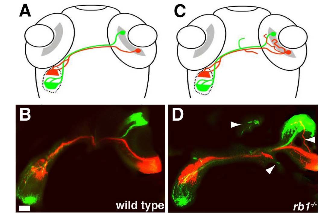
Fig. S3 Retinotopic mapping is intact in rb1te226a tectum. Dorsal views of schematized (A, C) and confocal projections (B, D) of retinotectal projection in wild type (A, B) and rb1te226a larvae at 120 hpf. DiO (green) labeled axons from anterior RGCs innervate the caudal tectum, while DiI (red) labeled axons of posterior RGCs project to the rostral tectum in both wild type and rb1te226a larvae. Arrowheads mark misprojecting axons. Scale bar = 50 μm.
Figure Data
Acknowledgments
This image is the copyrighted work of the attributed author or publisher, and
ZFIN has permission only to display this image to its users.
Additional permissions should be obtained from the applicable author or publisher of the image.
Full text @ PLoS Genet.